Fig. 1: Retracted pre-op smile view.
Fig. 2: Retracted pre-op frontal view.
Fig. 3: CBCT scan obtained using the CS 8100 3D.
Fig. 4: Pre-op bite registration with Futar.
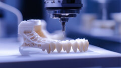
Fig. 5: Immediate dentures.
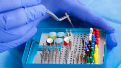
Fig. 6: Physics Forceps used for extractions.
Fig. 7: Area grafted and sutured.
Fig. 8: Try-in of immediate denture.
Fig. 9: Chairside soft denture relining material.
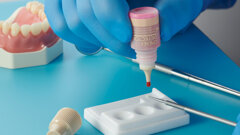
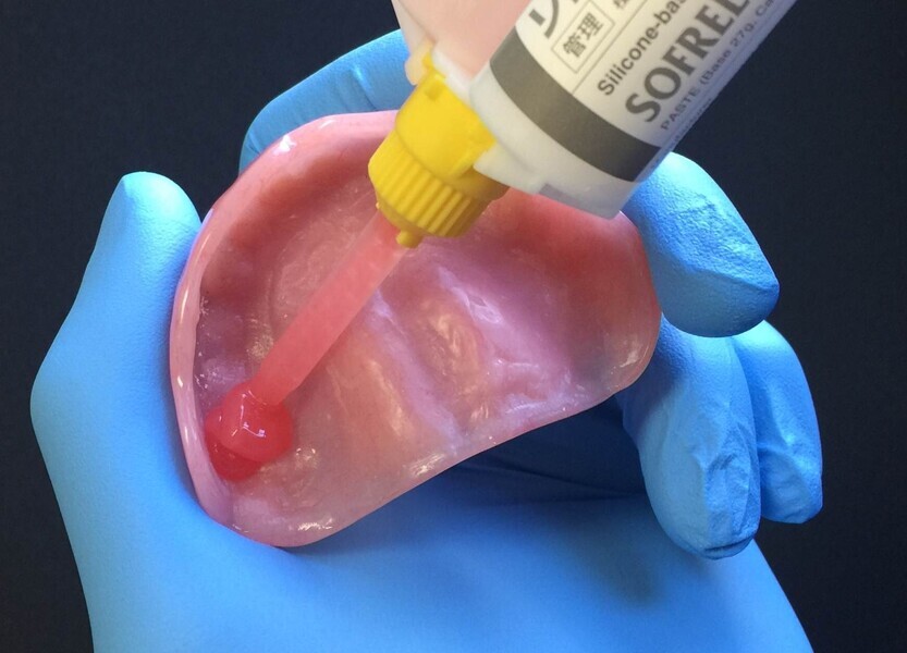
Fig. 10: Dispensing SOFRELINER TOUGH into immediate denture.
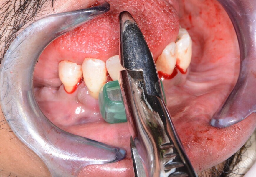
Fig. 11: Removing mandibular teeth.
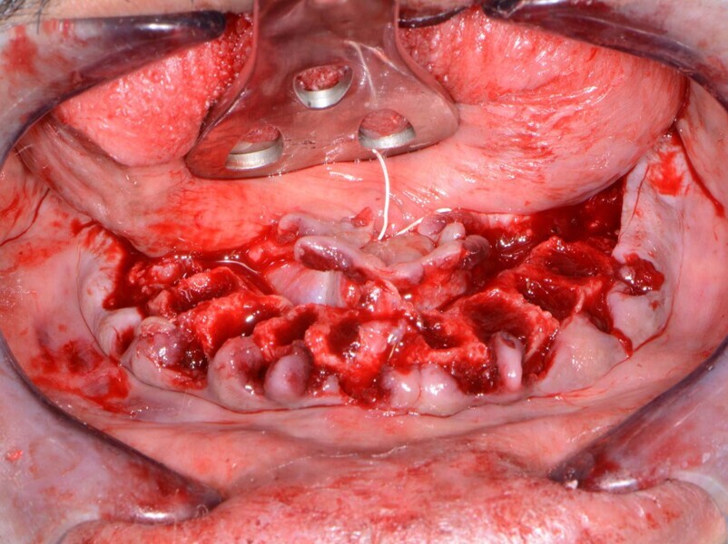
Fig. 12: Remaining ridge after tooth removal.
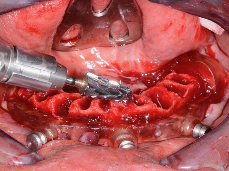
Fig. 13: Levelling bone using a CBCT-based guide.
Fig. 14: Positioning implant surgical guide.
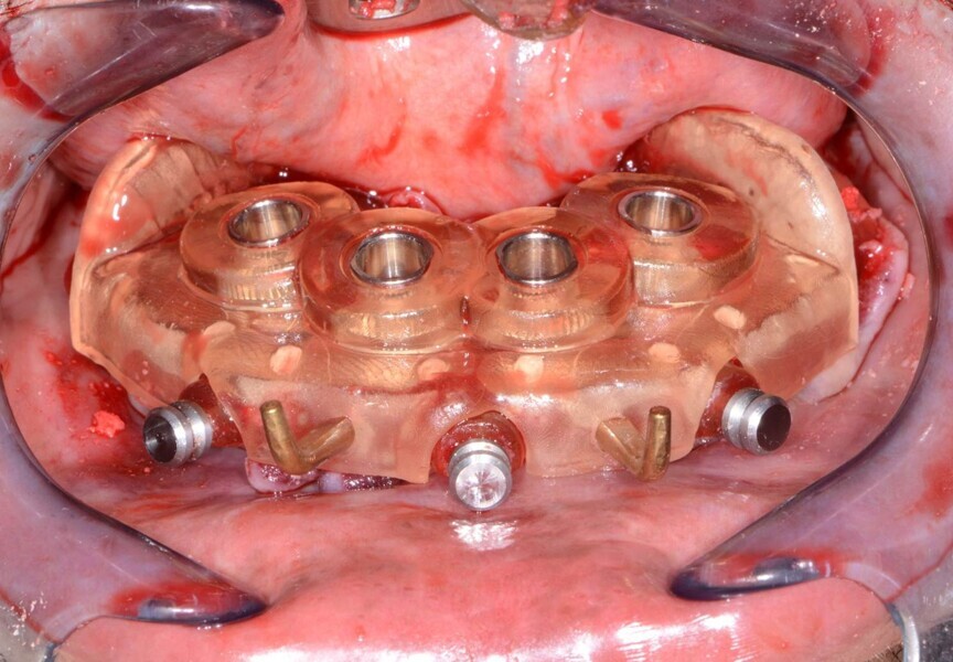
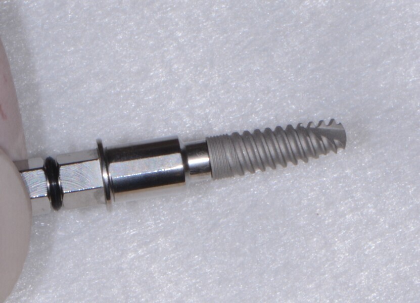
Fig. 15: Seven dental implant.
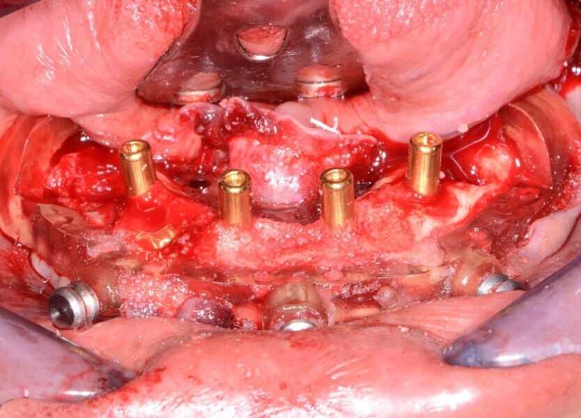
Fig. 16: Overdenture attachments inserted, followed by grafting material.
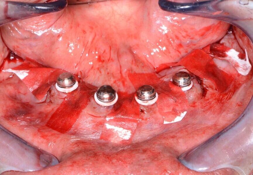
Fig. 17: Overdenture housings ready for the pick-up impression.
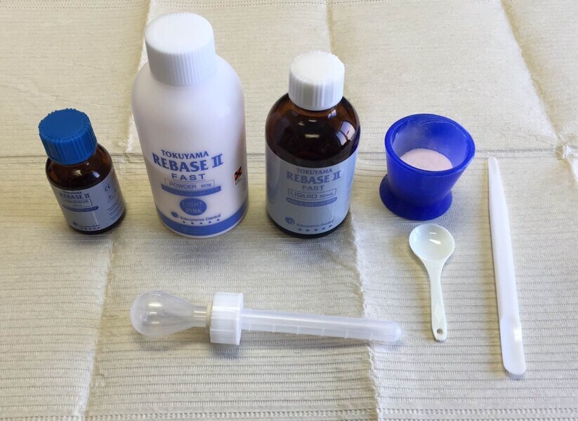
Fig. 18: Chairside hard denture relining material TOKUYAMA REBASE II.
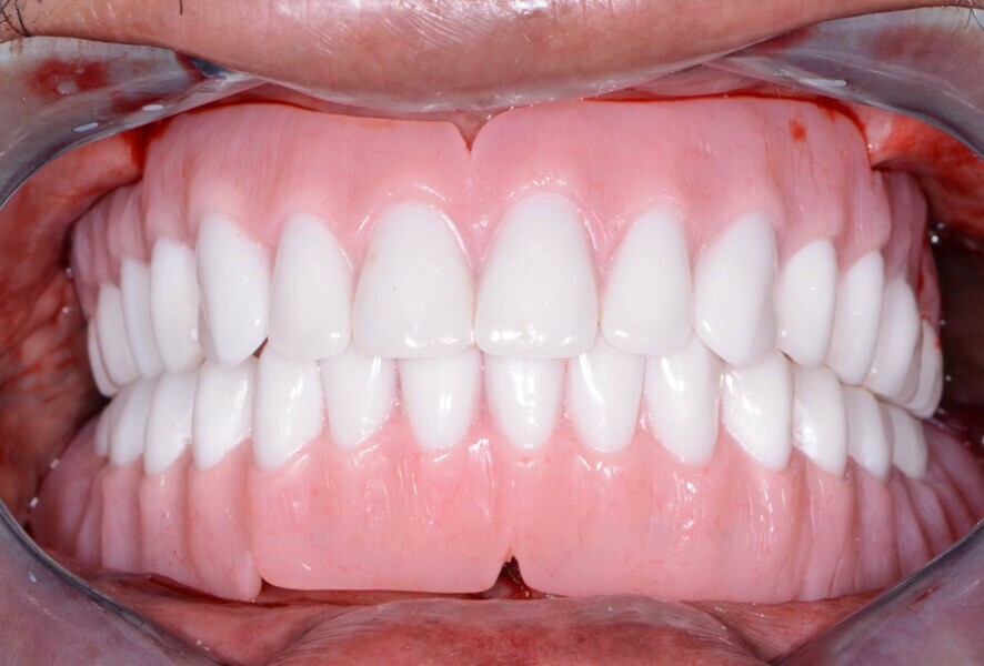
Fig. 19: Immediate post-op retracted view.
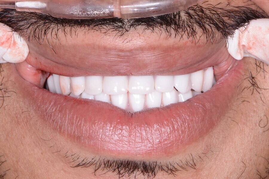
Fig. 20: Retracted post-op smile view.


 Austria / Österreich
 Bosnia and Herzegovina / Босна и Херцеговина
 Bulgaria / България
 Croatia / Hrvatska
 Czech Republic & Slovakia / Česká republika & Slovensko
 France / France
 Germany / Deutschland
 Greece / ΕΛΛΑΔΑ
 Hungary / Hungary
 Italy / Italia
 Netherlands / Nederland
 Nordic / Nordic
 Poland / Polska
 Portugal / Portugal
 Romania & Moldova / România & Moldova
 Slovenia / Slovenija
 Serbia & Montenegro / Србија и Црна Гора
 Spain / España
 Switzerland / Schweiz
 Turkey / Türkiye
 UK & Ireland / UK & Ireland
 Brazil / Brasil
 Canada / Canada
 Latin America / Latinoamérica
 USA / USA
 China / 中国
 India / भारत गणराज्य
 Pakistan / Pākistān
 Vietnam / Việt Nam
 ASEAN / ASEAN
 Israel / מְדִינַת יִשְׂרָאֵל
 Algeria, Morocco & Tunisia / الجزائر والمغرب وتونس
 Middle East / Middle East
