Owing to the introduction of magnification principles in dentistry, new techniques have been implemented for the successful performance of endodontic treatments. Microscopes have become an integral part of modern endodontics,1 and the use of conventional microscopy is becoming more frequent.2–4
Fig. 1: Alpha Air 6 dental operating microscope.
Despite the significant cost and training required, the use of the operating microscope is highly recommended to improve the visualisation of the operative field and to enhance the diagnostic capacity of the clinician, including the identification of isthmuses, accessory canals, complex pulp chamber anatomy, calcifications, obstructions and microfractures, among others,5, 6 which would otherwise be difficult to identify and treat. This results in better quality care and a higher success rate of treatments.7, 8 It has also been shown that the use of the operating microscope leads to a considerable improvement in ergonomics and therefore tends to reduce the occurrence of injuries related to poor posture and stress due to repetitive movements during the clinical workflow. All the advantages begin to be more palpable and applicable after going through the appropriate clinical training for acquiring the required skills to work under the microscope.9, 10
Through technological advances, a new generation of microscopic equipment with 3D technology has been developed that eliminates the binocular elements and offers an improvement in perception, clarity, depth of field, freedom of movement and clinical productivity in treatments. However, 3D microscopes have not been widely investigated, and scientific findings on their use and their influence on the fine motor skills of the operator are still limited. The purpose of this study was to evaluate and compare fine motor skills with the use of the conventional microscope and the 3D microscope in endodontic practice.
Methodology
Fifteen dentists who had no regular or recent clinical experience in the use of the operating microscope participated in this study. The study participants were final-year students and lecturers at the dental school of the Universidad Mariano Gálvez de Guatemala in Guatemala City. Each participant performed three manual tests of precision and dexterity divided into three stages as follows: unaided vision, using an Alpha Air 6 operating microscope (Seiler Instrument; Fig. 1) set to 8× magnification and using a PromiseVision 3D microscope (Seiler Instrument; Fig. 2) set to 8× magnification.
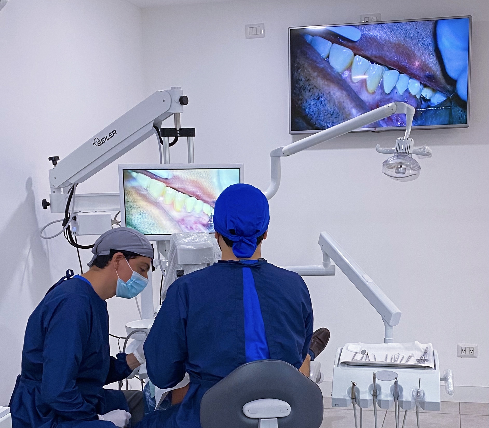
Fig. 2: The PromiseVision 3D surgical microscope being used during an apical microsurgery procedure.
All the dentists involved in the study received 6 hours of theoretical and practical training on the basic use of conventional and 3D microscopy. The training was given by second-year residents in endodontics from the Universidad Nacional Pedro Henriquez Ureña in Santo Domingo in the Dominican Republic. After the training, the participants performed the manual dexterity and fine motor skills tests. The tests required accurately penetrating a series of millimetric circular targets using a 21 mm #10 K-file. The targets were printed on a #20 calibre paper sheet with eight spaces, each space having ten circular targets inside (Fig. 3). Four of the spaces contained targets of 0.3 mm in diameter and the remaining four contained 0.35 mm diameter targets, corresponding to the letter “O” calibration in sizes 2 and 2.5, respectively. The position of each target within the field was determined by a Microsoft Excel randomised number generator.
Fig. 3: Sheet chart used during the fine motor skills tests.
During the fine motor skills tests, the time that the participants took to complete the test was recorded, from the penetration of the first target to the penetration of the last target (Fig. 4). To score accuracy and dexterity, a grading system of 0–3 points was used, 0 being the least accurate and 3 being the most accurate. A score of 3 was assigned if the file penetration was entirely within the target, a score of 2 was recorded if the penetration touched the edge of the target and was more than 50% within the target, a score of 1 was assigned if the penetration touched the edge of the target, but was more than 50% off the target, and a score of 0 was assigned if the target was completely intact, was missed or was penetrated more than once.
The completed test sheets were evaluated by two calibrated blinded evaluators with the help of a tabletop microscope. The scores for the 80 targets were calculated individually, obtaining a maximum possible score of 240. The statistical analysis was performed using the RealStatistics Using Excel program. The Shapiro–Wilk p > 0.01 test was performed to evaluate the normality of the data sample.
Fig. 4: The study participants working on the fine motor skill tests.
Results
Using the one-factor analysis of variance test for correlated differences, statistically significantly lower precision (p < 0.05) was found for working without magnification. The Tukey post hoc test showed statistically significantly greater precision (p < 0.05) when the 3D microscope was used. The one-factor analysis of variance test and Tukey’s post hoc tests found statistically significant differences (p < 0.05) in terms of the time needed to perform the precision test, working without magnification taking less time than working with conventional magnification and with 3D microscopy.
The time needed for the operator to adjust the microscope and to feel comfortable to start working on the tests was also measured. A shorter adjustment time was needed when the 3D microscope was used in comparison with the conventional microscope, and this difference was statistically significant. The mean adjustment time was 1.19 and 4.13 minutes, respectively.
The analysis of the results revealed that the tests with the greatest difference (p < 0.05) in both variables (time required and precision demonstrated) were those carried out when working without magnification, compared with working with conventional and 3D microscopy. It can be seen in Table 1 that it took less time on average to perform the test when not using magnification, but the scoring results on accuracy were directly proportional to time: the less time it took to perform the test, the less accurate the operator was. With the use of conventional and 3D magnification, significant differences were found in both time and effectiveness.
It can be seen in Table 1 that the completion time was shorter for the tests using the 3D microscope, compared with the tests carried out with a conventional microscope. The accuracy score obtained was higher when the conventional microscope was used. It is worth mentioning that the precision tests were performed on flat images, which may have influenced the perception of objects when performing the test using the 3D microscope. It is recommended to carry out a similar study by carrying out precision tests on 3D objects.
Table 1: Time differences and effectiveness
Average time to complete the tests in seconds (CI)
Mean microscopic precision score (CI)
No magnification
304 (259–347)
150 (128–171)
Conventional microscope
656 (525–780)
193 (173–210)
3D microscope
640 (554–773)
185 (174–197)
Discussion
It is necessary to understand the importance of magnification to achieve quality results in dental procedures and to reduce the margin of error, and the use of magnification in turn requires fine motor skills in dentistry. That is why this study aimed to evaluate and compare fine motor skills without the use of magnification, with the use of a conventional microscope and with the use of a 3D microscope. Over the years, the advantages of magnification in dentistry have been demonstrated. Now, we have progressed to studying the new 3D magnification system plus the contributions that it can make to clinical practice.
The results showed that the magnification systems used effected an increase in the fine motor skills of the participants, regardless of the type of magnification used. Regardless of the time it took to learn to work under the microscope or to complete a test, it is evident that the use of magnification improved the results and made the motor skills of the participants more efficient, resulting in marked precision during the testing. These results are quite similar to those reported by Wajngarten et al., demonstrating that magnification makes a significant contribution to and allows for better results in clinical work.7, 11, 12
It is understandable that, initially, the working time tends to be shorter when the microscope is not used, and the quality of the work is directly proportional to the time needed to perform the task. Using magnification requires theoretical and practical learning that, once achieved, will provide advantages in quality of work and improvements in the operator’s motor skills and ergonomics.7, 8, 13 It is important to note, as previously mentioned, that the use of magnification provides better visualisation and illumination of the operative field, helps to avoid long-term health problems, reduces the probabilities of occupational stress and improves working position.9, 10 The contributions of conventional microscopy have been well studied. It has effected a positive change in modern dentistry, facilitating better quality treatments with less execution time and higher success rates and thereby promoting a more pleasant experience for the dental professional and for the patient.
Despite being a relatively new magnification device and little studied so far, the 3D microscope achieves the desired quality standards. It is a tool that makes it easier for us to achieve results like those obtained with conventional microscopy and has the additional advantage of offering greater freedom of working position to the operator and an outstanding depth of field. Regarding the comparison with conventional microscopy, some differences could be linked to the time it takes to master the use of this technology; however, both magnification tools provide a considerable contribution to the execution of any dental treatment.13, 14
Conclusion
Through evaluating and comparing fine motor skills with the conventional microscope and the 3D microscope, we found that both devices contributed to the enhancement of fine motor skills, allowing the participants to achieve better results. 3D microscopy is a novel tool that is likely to become part of the standard equipment in dentistry, contributing positively to the implementation of microscopy in all specialties of dentistry.
MILAN, Italy: The 2026 International Symposium on Dental Hygiene (ISDH) is bringing together dental hygienists, clinicians, educators, researchers and other...
DUBAI, UAE: Dubai Dental Week, originally scheduled to take place from 16 to 21 January 2027, has been postponed to 2028. The organisers said that strong ...
SEOUL, South Korea: In June, dental professionals from across Europe visited Seoul to attend the 2026 Osstem Europe Tour Seminar. Organised by Osstem ...
Ahead of the 2026 Osstem Europe Meeting in Prague in the Czech Republic on 13–14 November, Dental Tribune International spoke with Dr Ieva Gendvilienė,...
SEOUL, South Korea: Osstem Implant invests heavily in research and development to strengthen its technological competitiveness, particularly in digitally ...
SEOUL, South Korea: Dental implant manufacturer Osstem Implant is expanding its corporate social responsibility (CSR) activities worldwide. Guided by its ...
BANGKOK, Thailand: Held on 27 and 28 March in the Thai capital, the Osstem World Meeting 2026 Bangkok highlighted the latest clinical strategies and ...
Prof. Adam Nulty is a leading expert in digital implant workflows. At IDEM 2026 in Singapore, he will challenge a common assumption in all-on-X dentistry: ...
SEOUL, South Korea: The Osstem Master Course is a comprehensive, integrated clinical programme comprising 24 sessions. Moving beyond one-off courses, it ...
SEOUL, South Korea: Osstem Implant has published a consensus paper after convening internationally recognised experts to discuss clinical factors affecting ...
SEOUL, South Korea: Osstem Implant has outlined the scale of its global manufacturing network and quality management system as part of its strategy to ...
Education
Live webinar Mon. 13 July 2026 11:30 am EST (New York)

 Brazil / Brasil
 Canada / Canada
 Latin America / Latinoamérica
 USA / USA
 Austria / Österreich
 Bosnia and Herzegovina / Босна и Херцеговина
 Bulgaria / България
 Croatia / Hrvatska
 Czech Republic & Slovakia / Česká republika & Slovensko
 France / France
 Germany / Deutschland
 Greece / ΕΛΛΑΔΑ
 Hungary / Hungary
 Italy / Italia
 Netherlands / Nederland
 Nordic / Nordic
 Poland / Polska
 Portugal / Portugal
 Romania & Moldova / România & Moldova
 Slovenia / Slovenija
 Serbia & Montenegro / Србија и Црна Гора
 Spain / España
 Switzerland / Schweiz
 Turkey / Türkiye
 UK & Ireland / UK & Ireland
 China / 中国
 India / भारत गणराज्य
 Pakistan / Pākistān
 Vietnam / Việt Nam
 ASEAN / ASEAN
 Israel / מְדִינַת יִשְׂרָאֵל
 Algeria, Morocco & Tunisia / الجزائر والمغرب وتونس
 Middle East / Middle East

 Dr. Fernando FranchLive webinar
 Dr. Nicolas OuelletRegister now1CELive webinar

 Dr. Nisha D’Silva BDS, MSD, PhD, Dr. Kıvanç Bektaş-KayhanRegister now1CELive webinar
 Federico ZunicaRegister now1CE


To post a reply please login or register