Search Dental Tribune

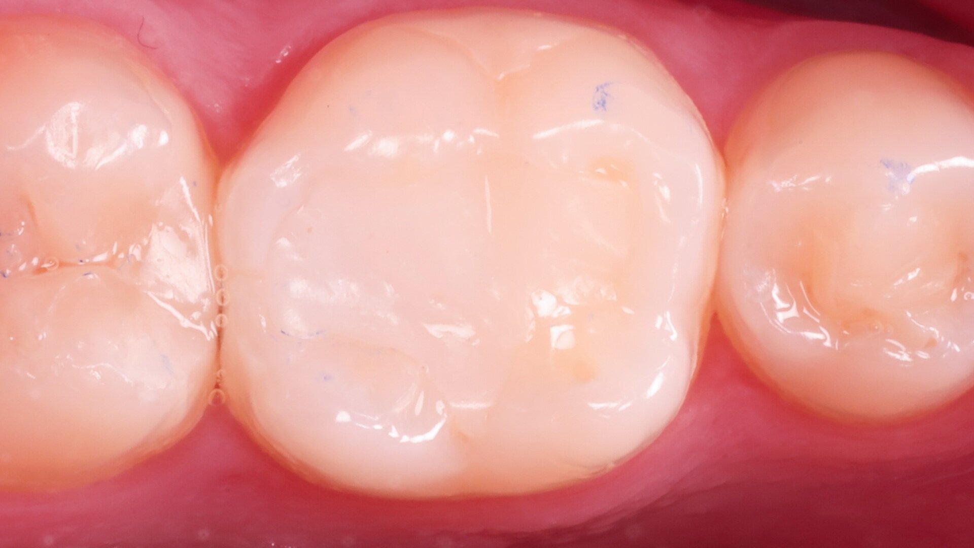
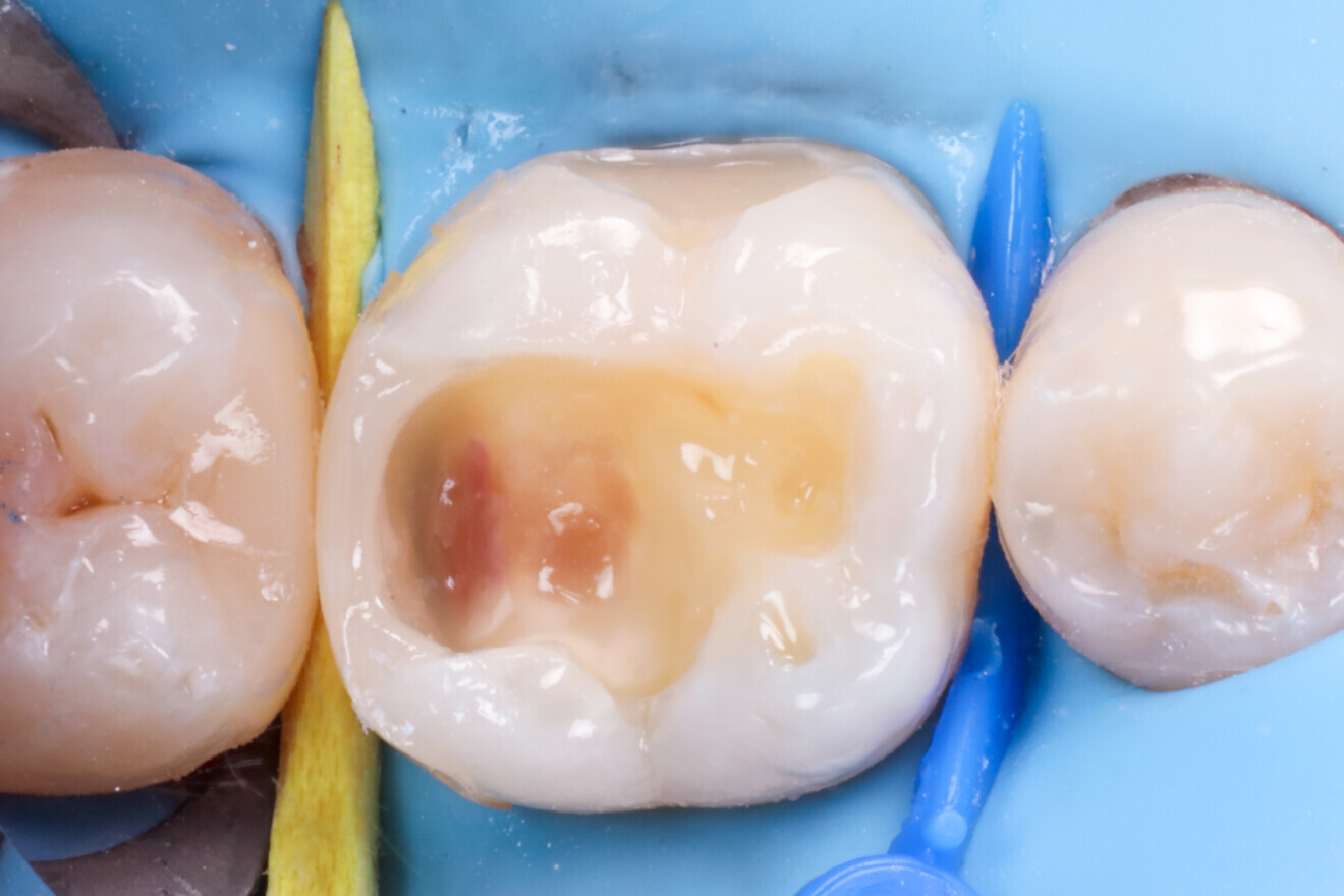
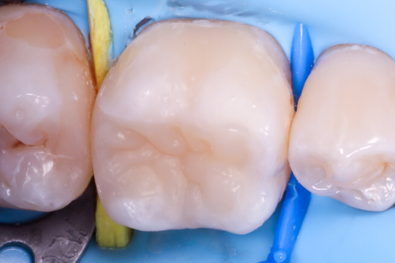
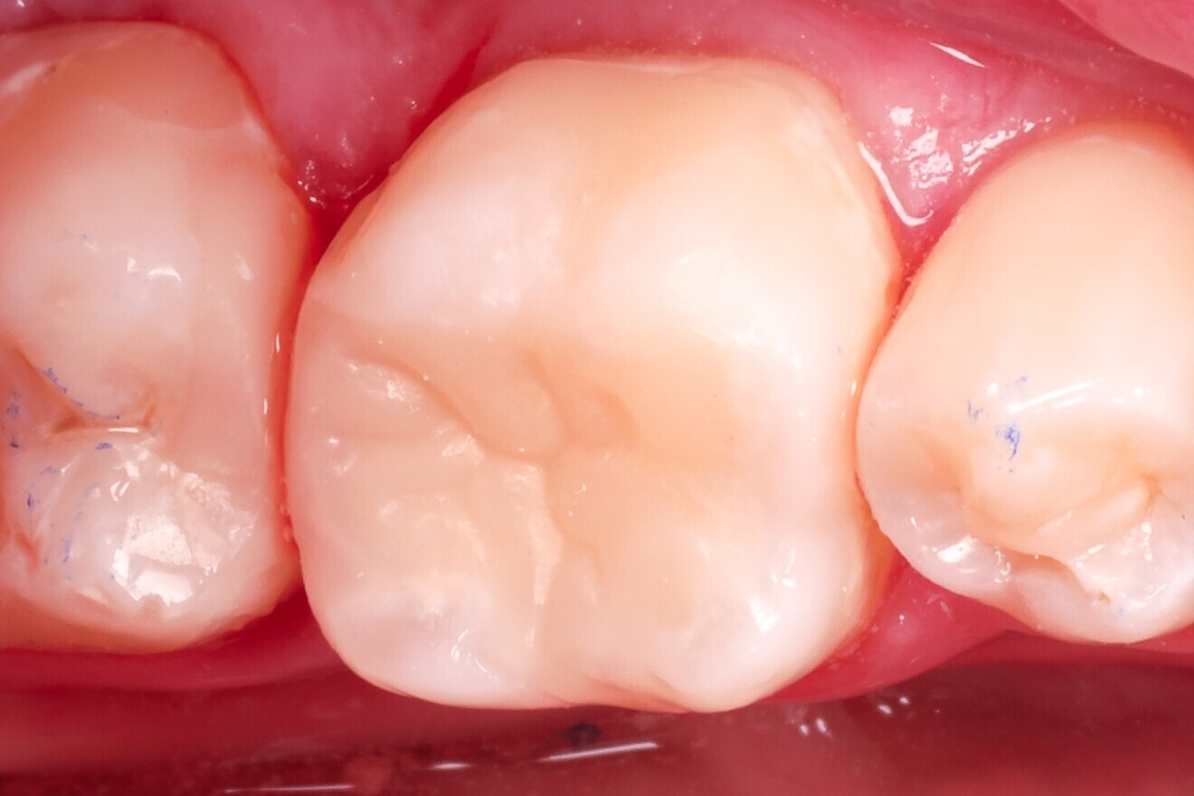
Fig. 1: Pre-op view of the mandibular first molar with existing composite restorations. (All images: Dr Péter Farkas)

Mon. 6. October 2025

save

In recent years, manufacturers have developed increasingly advanced composite materials, offering improvements in aesthetics, handling and mechanical properties. However, none of these innovations have truly eliminated the challenge of polymerisation shrinkage. This stress, caused by volumetric shrinkage during polymerisation, generates internal stress within the restoration, which can weaken adhesive bond strength, cause marginal gaps and result in postoperative sensitivity or secondary caries.

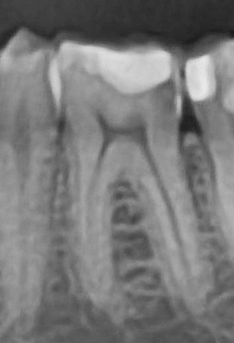
Fig. 2: Panoramic radiograph showing a distal carious lesion.

Fig. 2: Panoramic radiograph showing a distal carious lesion.

The more clinicians have learned about adhesion and composite behaviour, the more cautious they have become with layering techniques to ensure long-term success. Unfortunately, these cautious techniques—especially when aiming to reduce stress—often require significantly more chairside time. There is a growing need for a method that simplifies layering while preserving adhesive integrity and aesthetic outcomes.

Indirect restorations have proved to be highly effective in terms of adhesion, particularly because they allow the adhesive interface to mature fully before the restoration is bonded under minimal stress. This is especially true when the preparation is immediately followed by adhesive treatment—known as immediate dentine sealing. In this approach, freshly cut dentine is sealed right after tooth preparation, allowing the resin-infiltrated dentine layer, or hybrid layer, to mature in a stress-free environment during the laboratory phase of restoration fabrication. Since no shrinkage-inducing composite is placed at this stage, the adhesive layer remains undisturbed and can fully develop its mechanical properties before final bonding.

However, these indirect approaches are not always feasible in everyday clinical settings owing to time, cost or patient preference. This has led to increased interest in so-called semi-direct techniques, an approach that blends aspects of both direct and indirect workflows. The term “semi-direct” refers to restorations that are either fabricated outside the mouth on a model created from a quick impression or built directly on the tooth using a separating material such as PTFE tape. In both scenarios, the restoration undergoes extra-oral polymerisation before being returned and bonded to the tooth. This approach allows for improved control over polymerisation stress while preserving the efficiency and practicality of a chairside procedure. Although semi-direct methods offer great potential, many existing techniques are complex and demand artistic skills for free-hand anatomical shaping, limiting their accessibility in daily practice.

The SEAL (Stress-reduced, Esthetic, Anatomically guided, Layer-less) concept is a simplified semi-direct approach that eliminates the need for artistic modelling. Instead, it uses a preformed anatomical model, allowing clinicians to transfer morphology in a copy–paste fashion. The following case illustrates the application of the SEAL concept.

Technique description

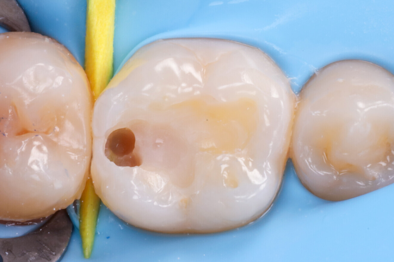
Treatment focused on a mandibular first molar with existing Class I and V composite restorations (Fig. 1). The tooth was asymptomatic, and cold testing revealed a normal response. A distal carious lesion, which was not clinically visible, was first suspected during routine screening and its extent clearly identified on the panoramic radiograph (Fig. 2).

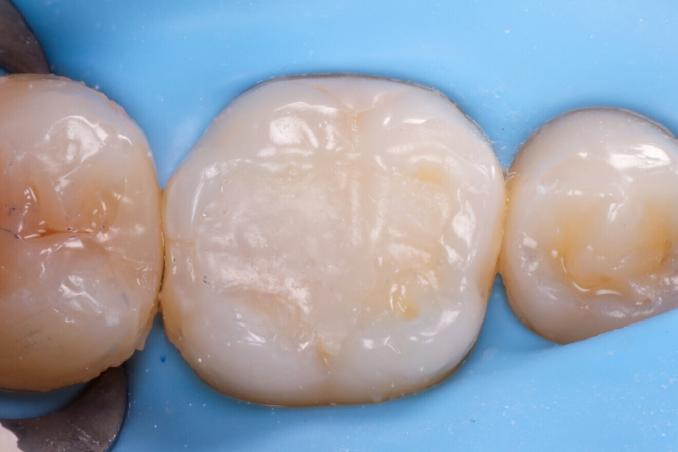
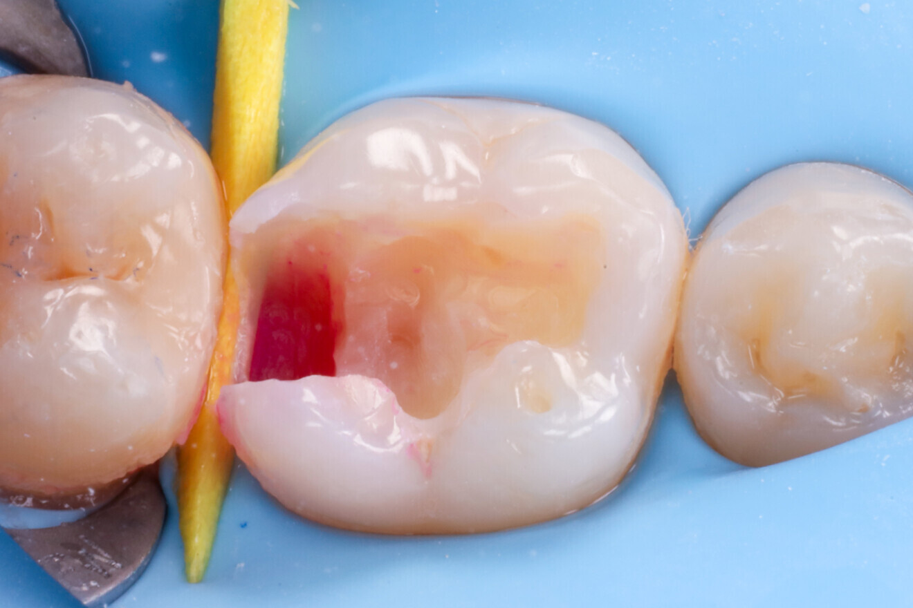
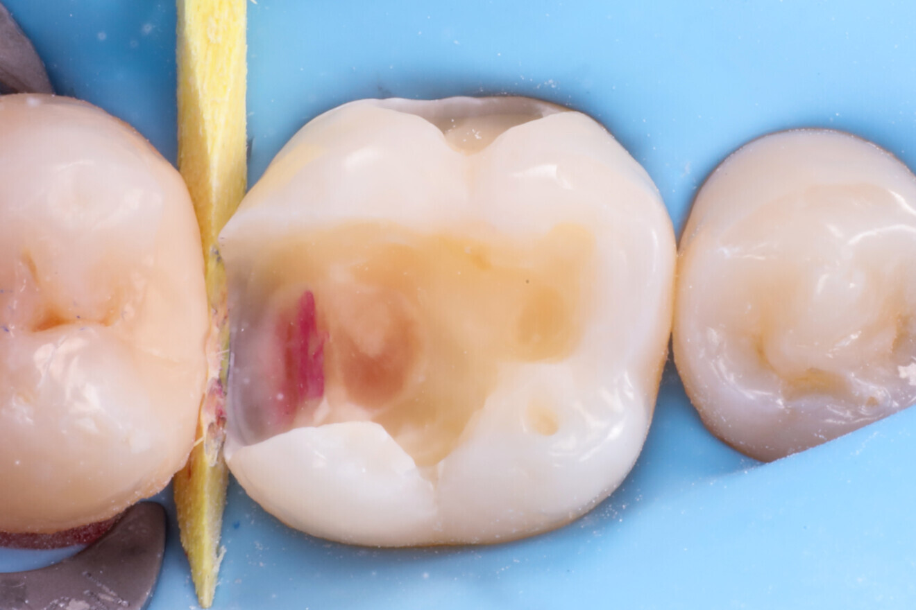
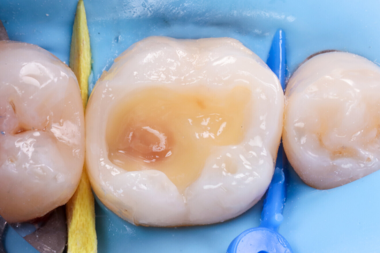
After isolation using a dental dam (Isodam, 4D Rubber; Fig. 3), the old restorations were removed, revealing the distal carious lesion (Fig. 4). Caries detection dye (CARIES DETECTOR, Kuraray Noritake Dental) was applied for visual confirmation (Fig. 5).

Fig. 3: Dental dam isolation.

Fig. 3: Dental dam isolation.

Fig. 4: Distal lesion becoming visible during composite removal.

Fig. 4: Distal lesion becoming visible during composite removal.

Fig. 5: Caries detection dye applied to visualise the extent of the carious lesion.

Fig. 5: Caries detection dye applied to visualise the extent of the carious lesion.

After removal of the decayed tissue, the cavity was air-abraded with 29 µm aluminium oxide at 200 kPa pressure using an air abrasion system (AquaCare, Velopex International; Fig. 6).

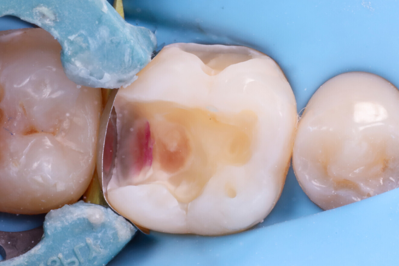
A double-curved sectional matrix (TOR) was placed with a universal ring (Palodent V3, Dentsply Sirona; Fig. 7). The enamel was etched for 20 seconds with a 36% phosphoric acid (BLUE ETCH, Cerkamed), rinsed and dried completely. The whole cavity was treated using a gold standard two-step self-etch adhesive system (CLEARFIL SE Protect, Kuraray Noritake Dental), followed by light polymerisation for 20 seconds. A thin layer (~0.5 mm) of a flowable composite (Estelite Universal Flow, High; Tokuyama Dental) was applied and polymerised for another 20 seconds using a 1,500 mW/cm² curing light (Curing Pen, Eighteeth).

The proximal wall was rebuilt incrementally with a paste composite (Estelite Asteria, Shade NE), each layer polymerised for 10 seconds (Fig. 8). Undercuts were filled with a short-fibre-reinforced flowable composite (everX Flow, GC) in increments polymerised for 20 seconds (Fig. 9).

Fig. 6: Cavity after air abrasion.

Fig. 6: Cavity after air abrasion.

Fig. 7: Sectional matrix system in place.

Fig. 7: Sectional matrix system in place.

Fig. 8: Proximal wall built incrementally with composite.

Fig. 8: Proximal wall built incrementally with composite.

Fig. 9: Undercuts filled with a short-fibre-reinforced composite.

Fig. 9: Undercuts filled with a short-fibre-reinforced composite.

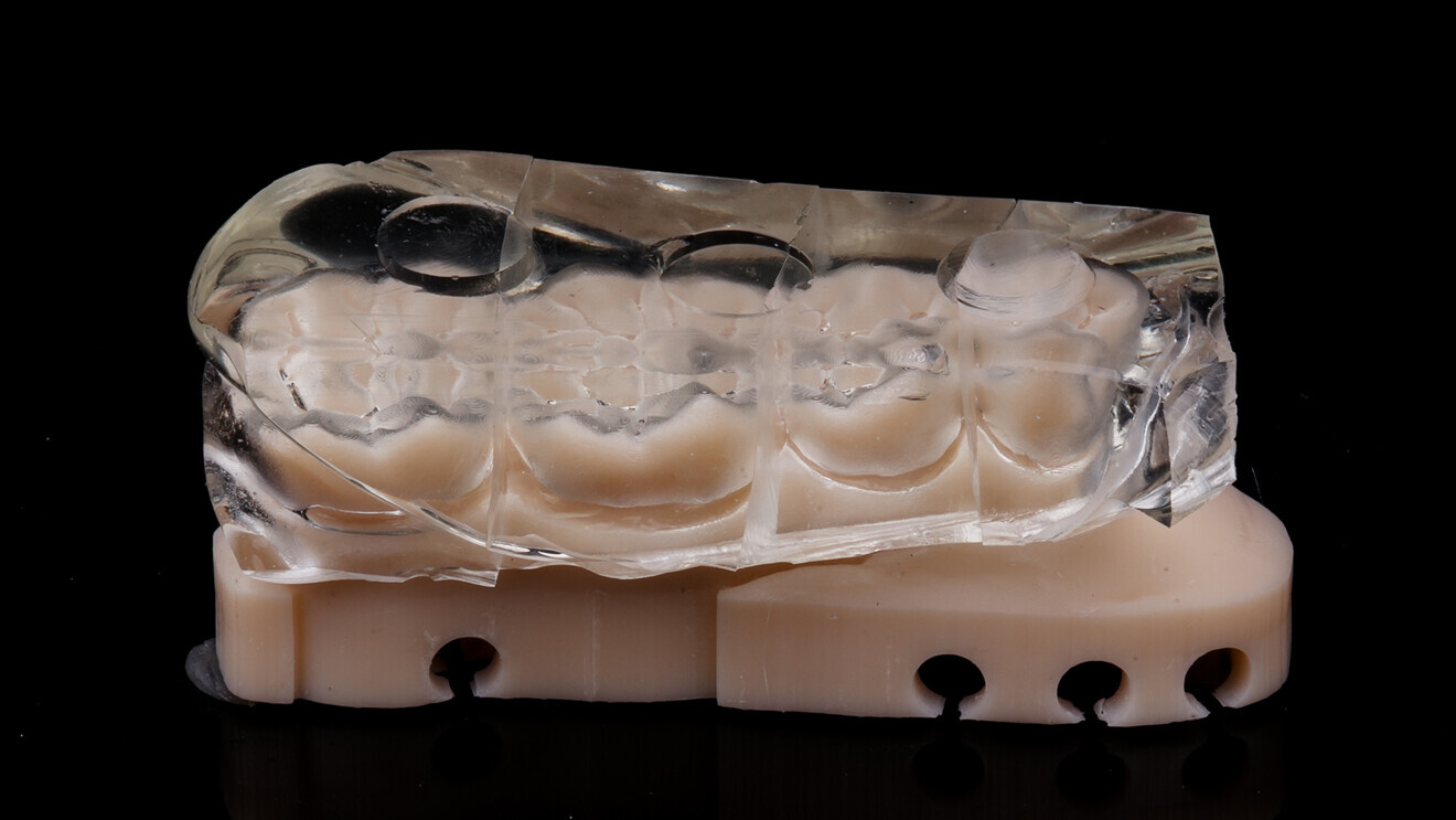
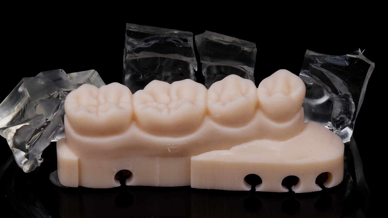
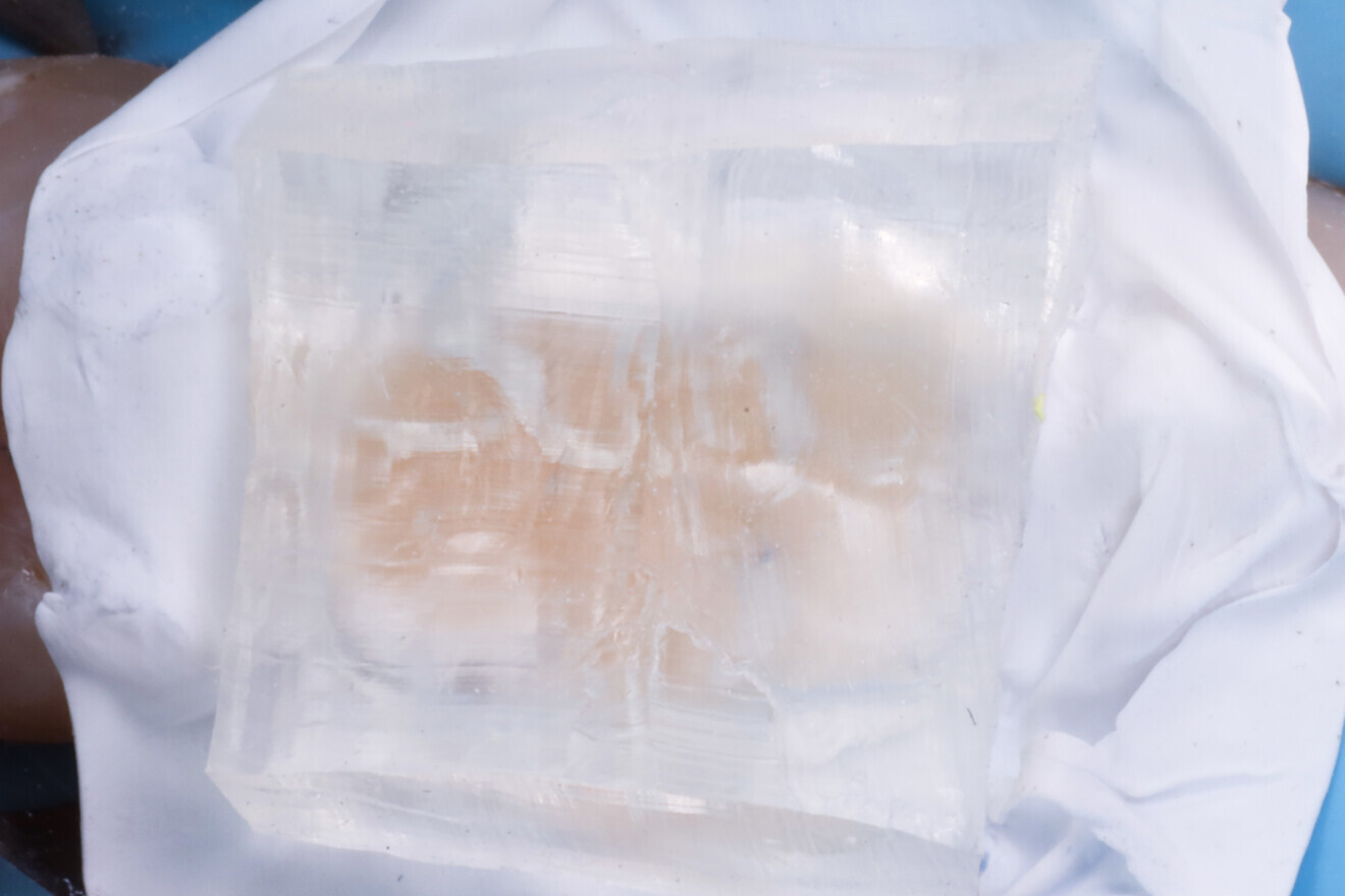
To perform the occlusal copy–paste technique, a preformed anatomical diagnostic 3D-printed model created by master dental technician János Makó was used. A clear silicone index (EXACLEAR, GC) was fabricated from this model (Fig. 10). The index was sectioned to isolate the area corresponding to the first molar (Fig. 11) and trimmed to shape for easier intra-oral adaptation (Fig. 12). The silicone index was then separated from the printed model, ready for clinical use (Fig. 13).
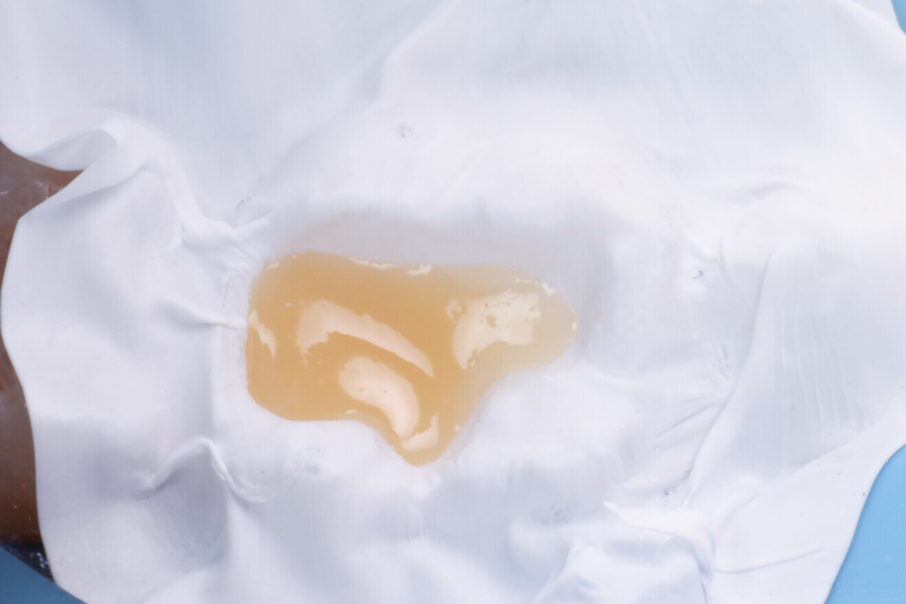
Fig. 10: 3D-printed model and clear silicone index.

Fig. 10: 3D-printed model and clear silicone index.

Fig. 11: Silicone index sectioned tooth by tooth.

Fig. 11: Silicone index sectioned tooth by tooth.

Fig. 12: Silicone index sectioned for the first molar.
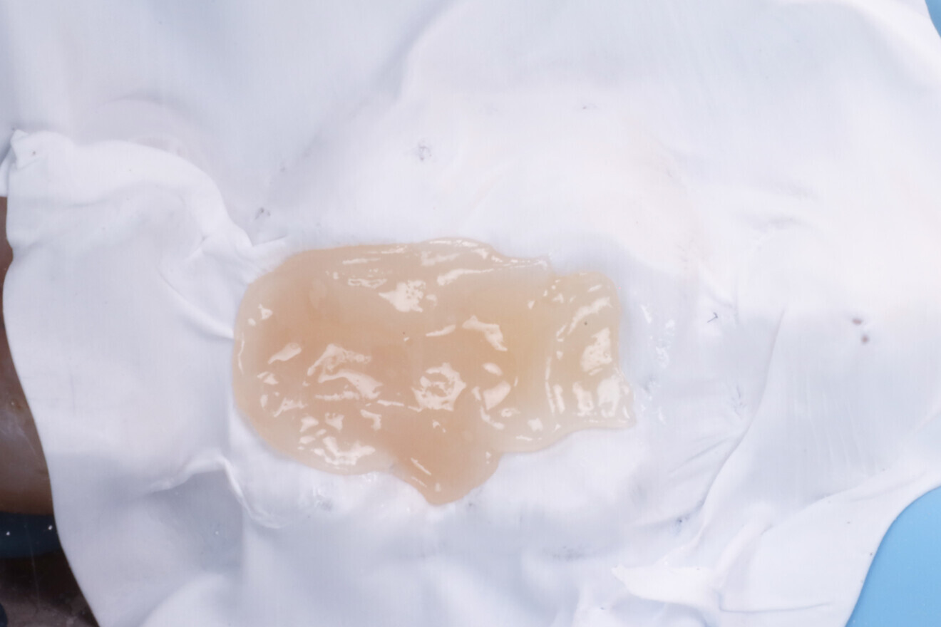
Fig. 12: Silicone index sectioned for the first molar.

Fig. 13: Final clear silicone index ready for intra-oral use.

Fig. 13: Final clear silicone index ready for intra-oral use.

Fig. 14: PTFE tape separating the cavity surface to prevent bonding with the composite.

Fig. 14: PTFE tape separating the cavity surface to prevent bonding with the composite.

Fig. 15: Short-fibre-reinforced composite placed in the dentine region.

Fig. 15: Short-fibre-reinforced composite placed in the dentine region.

Fig. 16: Occlusal enamel composite before polymerisation.

Fig. 16: Occlusal enamel composite before polymerisation.

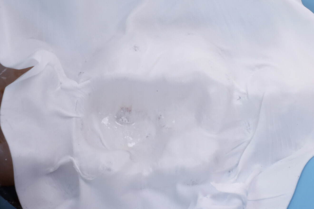
Fig. 17: Silicone index seated over the unpolymerised composite.

Fig. 17: Silicone index seated over the unpolymerised composite.

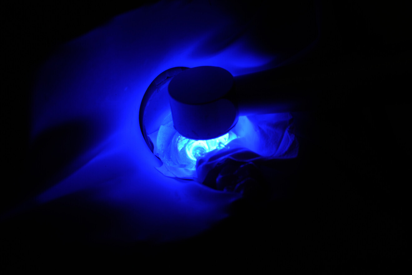
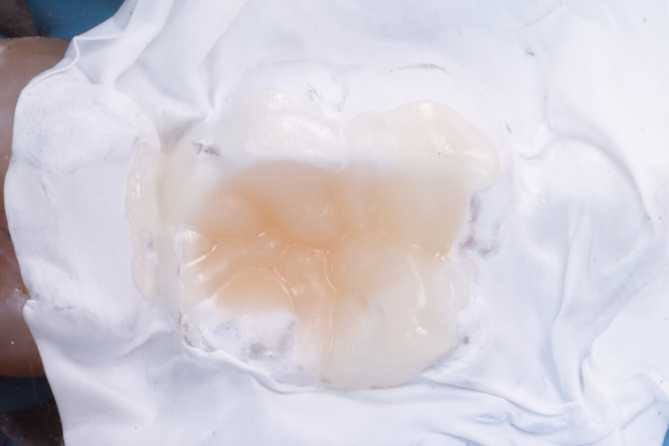
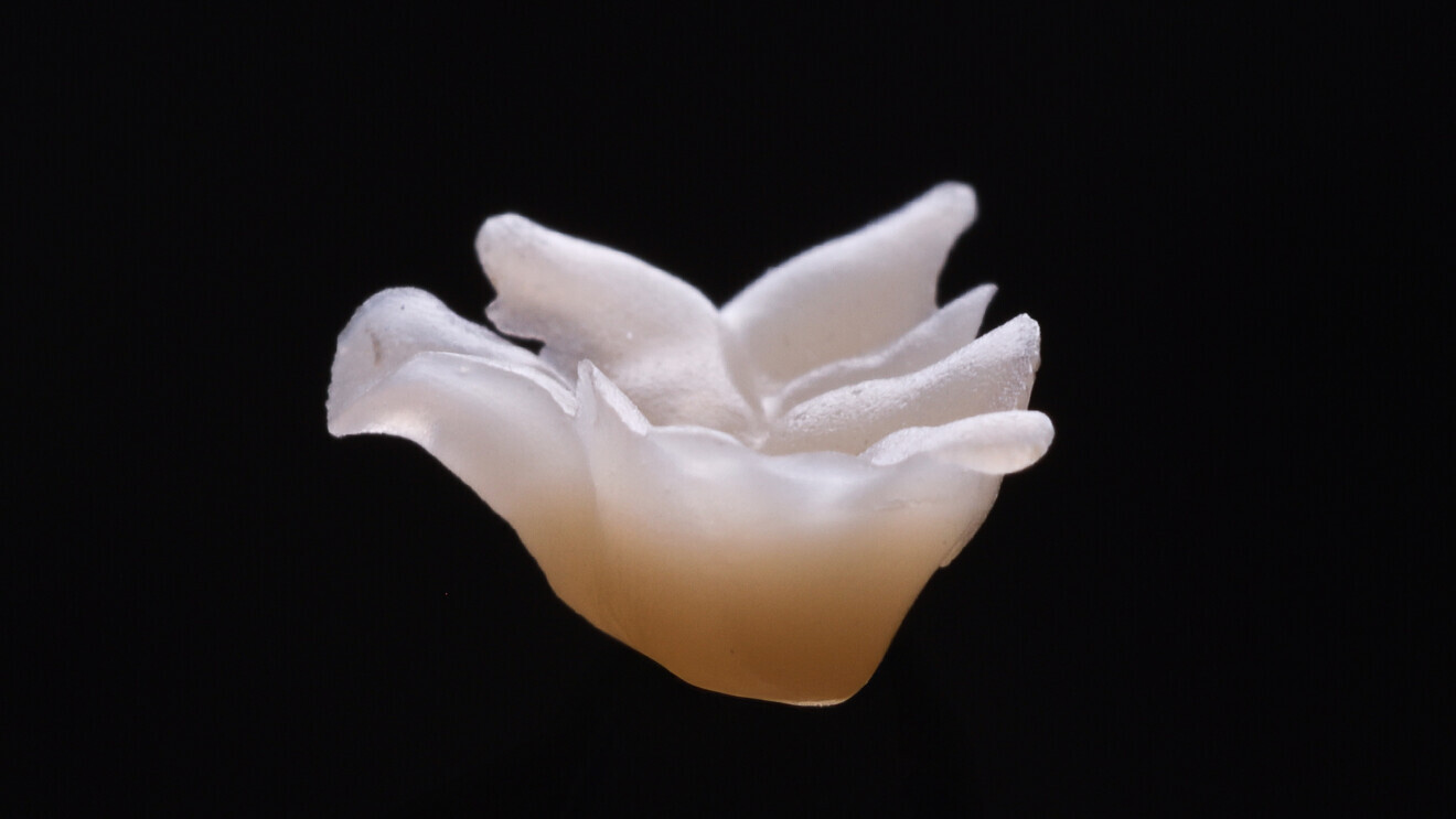
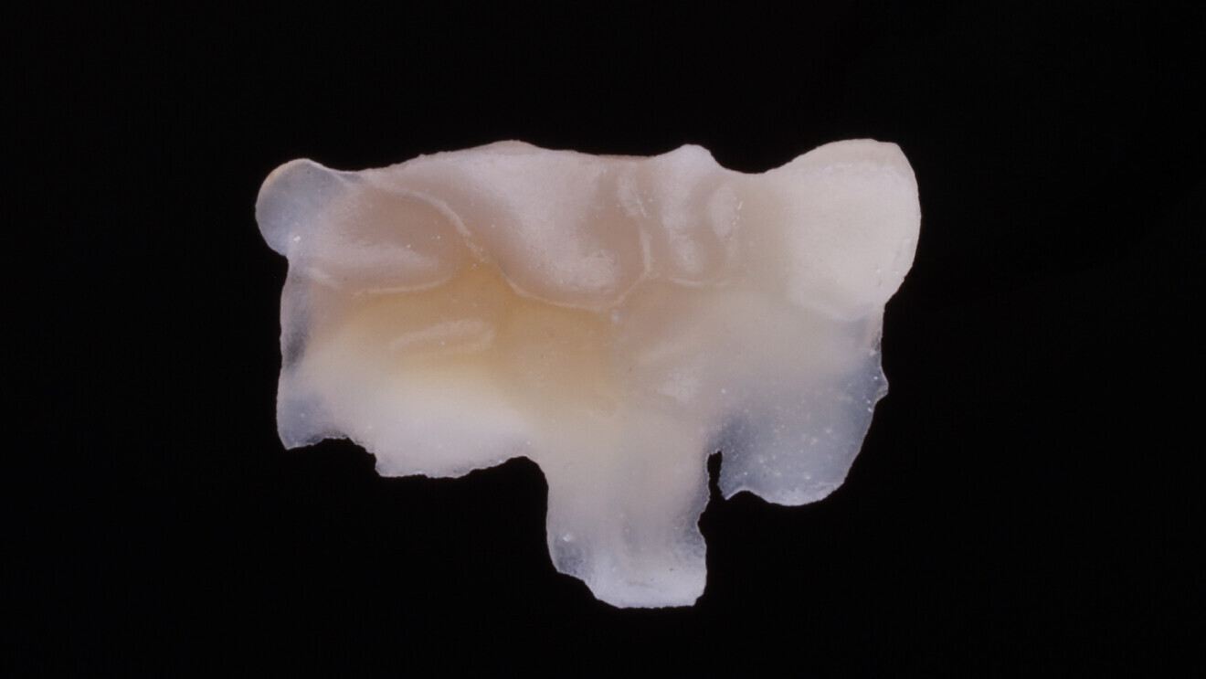
The cavity was then covered with PTFE tape to prevent bonding between the composite layers and the cavity surface (Fig. 14), and everX Flow was applied in the dentine region and light-polymerised for 10 seconds (Fig. 15). This was followed by a layer of Estelite Asteria (Shade OcE), placed without initial polymerisation (Fig. 16). The anatomical silicone index was then placed over it with gentle pressure and light-polymerised (Figs. 17 & 18). After removing the silicone index, additional polymerisation was performed (Fig. 19). Extra-oral polymerisation followed, 20 seconds per side (Figs. 20 & 21).

Fig. 18: Light polymerisation through the silicone index.

Fig. 18: Light polymerisation through the silicone index.

Fig. 19: Composite after removal of the silicone index.

Fig. 19: Composite after removal of the silicone index.

Fig. 20: Extra-oral approximal view of the semi-direct inlay.

Fig. 20: Extra-oral approximal view of the semi-direct inlay.

Fig. 21: Extra-oral occlusal view of the semi-direct inlay.

Fig. 21: Extra-oral occlusal view of the semi-direct inlay.

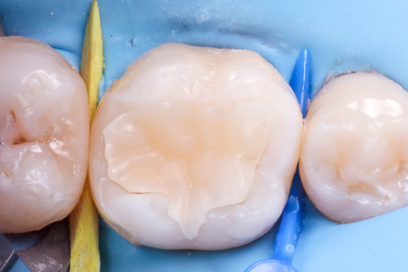
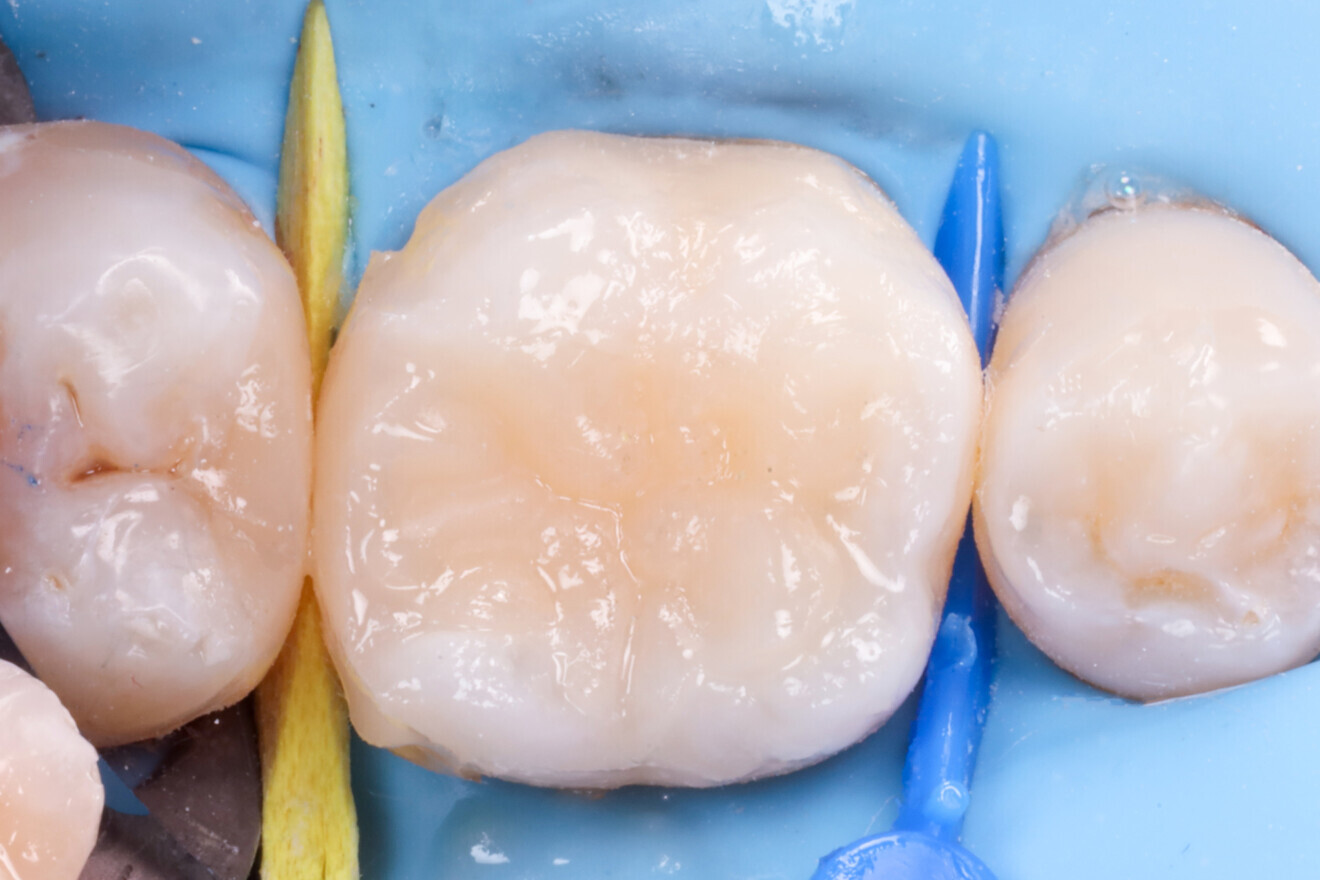
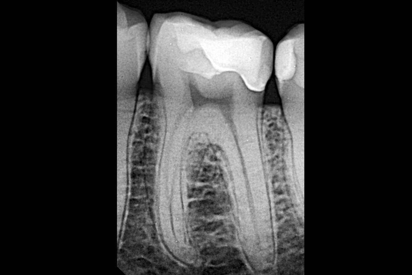
After try-in (Fig. 22), the restoration was bonded using a low-flow, highly filled composite (Estelite Universal Flow, SuperLow; Fig. 23). Excess material was removed (Fig. 24), followed by another round of light polymerisation for 20 seconds per side. Final excess removal was done (Fig. 25), and thanks to the preformed anatomy, most of the morphological detail remained intact even after occlusal adjustment (Fig. 26). A control radiograph was taken (Fig. 27).

Fig. 22: Try-in of the semi-direct restoration.

Fig. 22: Try-in of the semi-direct restoration.

Fig. 23: Cementation with a flowable composite.

Fig. 23: Cementation with a flowable composite.

Fig. 24: Before excess removal.

Fig. 24: Before excess removal.

Fig. 25: After excess removal and polishing of the restoration.

Fig. 25: After excess removal and polishing of the restoration.

Fig. 26: Occlusal view after final adjustment.

Fig. 26: Occlusal view after final adjustment.

Fig. 27: Final radiograph of the restoration.

Fig. 27: Final radiograph of the restoration.

Discussion and conclusion

A significant portion of everyday dental practice involves replacing existing restorations owing to secondary caries, postoperative sensitivity or structural fractures. These common clinical situations prompt an important consideration: how much does polymerisation shrinkage contribute to these failures?

Conceptually, there is a delicate balance between adhesive bond strength and polymerisation stress. When bond strength is low and stress is high, debonding is likely. Even if the bond is strong, excessive stress can still cause internal tension, leading to issues such as cusp deflection or marginal failure.

Thus, long-term clinical success depends not only on optimising bond strength but also on minimising shrinkage-induced stress. The SEAL concept addresses both factors. By allowing the hybrid layer to mature without stress—through the use of a PTFE barrier that temporarily separates the composite from the bonded surface—the method mimics the immediate dentine sealing strategy used in indirect restorations.

Furthermore, it replaces complex layering with an efficient copy–paste strategy using a clear silicone index derived from an anatomical model. This creates a semi-direct restoration that is both aesthetic and predictable and requires minimal artistic skill. The technique is approachable, easy to learn and highly relevant to general practice.

Topics:
Tags:
To post a reply please login or register
advertisement
advertisement