In addition to intra-oral and panoramic radiographs,various visual techniques are available for endodontic treatment today. Above all, information obtained through the dental microscope has become essential.
“See better, do better” is a slogan in modern endodontics. The dental microscope is a wonderful tool for problem-solving in endodontics, for instance for the removal of broken instruments and root-filling materials, finding missed canals, perforation repair, diagnosis of tooth fractures, evaluation of marginal integrity of restorations, precise manipulation in periradicular surgery and deep dental caries, and confirmation of root-canal cleanliness. Yoshioka et al. (2002), for example, reported that the rate of detection of root-canal orifices under a microscope was significantly higher than the number detected with the naked eye. It was also found that surgical loupes were relatively ineffective compared with the microscope.
In addition, computed tomography (CT) is becoming increasingly popular among endodontists, particularly in the assessment of difficult cases and for problem-solving in endodontic treatment. Higher use (34.2 per cent) of CBCT was demonstrated by a recent webbased survey of active members of the American Association of Endodontists in the US and Canada (Dailey et al. 2010). Owing to its high radiation dosage, however, careful consideration is needed before taking CT images. Consequently, a project team from the Japanese Association for Dental Science presented a report in 2010 on the use of CT in dentistry, and a joint position statement by the American Association of Endodontists and American Academy of Oral and Maxillofacial Radiology was issued in February 2011. The combined use of the dental microscope and CT for apicectomy was approved as an advanced dental technology by the Ministry of Health, Labor and Welfare in Japan in 2007, and seven Japanese dental hospitals have been using the technology since 1 February 2013.
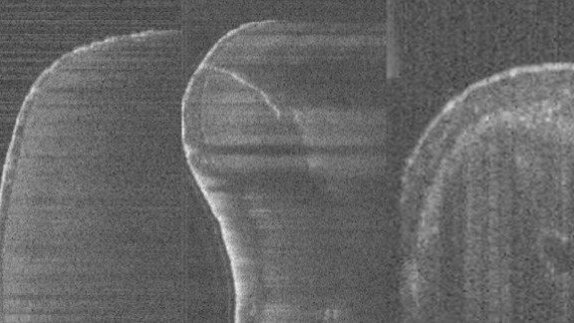
Optical coherence tomography (OCT) is a highresolution imaging technique that allows micro-metre-scale imaging of biological tissues over small distances. It was introduced in 1991 and uses infrared light waves that are reflected from the internal microstructure within the biological tissues (Shemesh et al. 2008). There have been reports on its use for intra-canal imaging, diagnosis of vertical root fracture (Yoshioka et al. 2013) and perforations. Since OCT isnon-invasive and free of radiation, this technology may be very useful for endodontic diagnosis and treatment (Figs. 1a–2).
This article was published in roots international magazine of endodontology No. 3/2013.
embedImagecenter("Imagecenter_1_973",973, "large");
Visual tools such as intra-oral scanning and photography have transformed the way clinicians communicate with their patients, making complex dental ...
How to achieve greater efficiency, safety and job satisfaction in endodontics—endodontist and trainer Dr Sabine Remensberger has been sharing her ...
In recent years, the technology associated with endodontic therapy has undergone a veritable revolution. For years, intraoral radiographs were used as the ...
This year, the International Dental Show’s (IDS) centenary year, the 40th edition of IDS, the world’s leading trade fair for dentistry, will focus on ...
COLOGNE, Germany: Align Technology recently announced that it was launching the iTero Element 5D imaging system, which provides a comprehensive approach to ...
LJUBLJANA, Slovenia: At this year’s International Dental Show (IDS), held from 25 to 29 March in Cologne in Germany, more than 135,000 visitors from 156 ...
GLATTFELDEN, Switzerland: On 16 and 17 June, COLTENE invited close to 100 dental experts from all over Europe to Zurich in Switzerland for a two-day seminar...
The technology of clear aligners has revolutionised modern orthodontics, offering an aesthetic and comfortable alternative to fixed appliances. Since its ...
After explaining the basic physics of the laser and its effects on both bacteria and dentinal surfaces, the second part of this article series will analyse ...
COLOGNE, Germany: Align Technology’s interactive booth at IDS 2021 represents the company’s largest-ever presence at the show. The company has ...
Live webinar
Tue. 10 February 2026
7:00 pm EST (New York)
Prof. Dr. Wael Att, Dr. Robert A. Levine DDS, FCPP, FISPPS, AOD, Dr. Larissa Bemquerer ITI Scholar at Harvard
Live webinar
Wed. 11 February 2026
11:00 am EST (New York)
Dr. med. dent. Sven Mühlemann
Live webinar
Wed. 11 February 2026
12:00 pm EST (New York)
Prof. Dr. Samir Abou Ayash
Live webinar
Fri. 13 February 2026
12:00 pm EST (New York)
Live webinar
Mon. 16 February 2026
12:00 pm EST (New York)
Live webinar
Tue. 17 February 2026
12:00 pm EST (New York)
Live webinar
Wed. 18 February 2026
9:00 am EST (New York)
Dr. Anna Lella, Ms. Francesca Nava


 Austria / Österreich
 Bosnia and Herzegovina / Босна и Херцеговина
 Bulgaria / България
 Croatia / Hrvatska
 Czech Republic & Slovakia / Česká republika & Slovensko
 France / France
 Germany / Deutschland
 Greece / ΕΛΛΑΔΑ
 Hungary / Hungary
 Italy / Italia
 Netherlands / Nederland
 Nordic / Nordic
 Poland / Polska
 Portugal / Portugal
 Romania & Moldova / România & Moldova
 Slovenia / Slovenija
 Serbia & Montenegro / Србија и Црна Гора
 Spain / España
 Switzerland / Schweiz
 Turkey / Türkiye
 UK & Ireland / UK & Ireland
 Brazil / Brasil
 Canada / Canada
 Latin America / Latinoamérica
 USA / USA
 China / 中国
 India / भारत गणराज्य
 Pakistan / Pākistān
 Vietnam / Việt Nam
 ASEAN / ASEAN
 Israel / מְדִינַת יִשְׂרָאֵל
 Algeria, Morocco & Tunisia / الجزائر والمغرب وتونس
 Middle East / Middle East











To post a reply please login or register