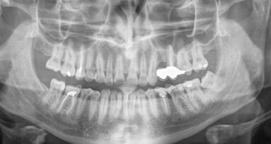
NAGOYA, Japan: With the aim of clarifying the relationship between panoramic radiographic appearance and longitudinal CBCT classification of root configurations of the mandibular second molar, researchers from Japan have investigated hundreds of panoramic radiographs and are now planning to develop an artificial intelligence (AI) system to help simplify diagnosis of mandibular second molar root canal configurations on panoramic radiographs in the future.
For the study, Drs Takuma Funakoshi and Takuya Shibata, who both work at the Department of Oral and Maxillofacial Radiology at the Aichi Gakuin University’s School of Dentistry, and their research team examined panoramic radiographs of 1,058 mandibular second molars and classified them into five types according to the number and configuration of the roots.
These molars were also examined with CBCT at four levels between the pulp chamber and the root apex, and axial images perpendicular to the root axis were categorised into three patterns:
- single (fused root with small grooves on both buccal and lingual sides or a round root with one canal);
- double (two separate roots with a trabecular appearance between them); and
- C-shaped (root with a deep groove opening only on the lingual or buccal side relative to the opposite side).
Based on these patterns and their scan levels, the CBCT root morphology appearance in each tooth unit was classified into one of seven groups. The scientists then investigated the relationship between these seven CBCT groups and the five panoramic root types.
It was found that, in panoramic Type 1 and 2 (with separate roots), 85% had roots with a double pattern (Groups II and III) on the CBCT images. In panoramic Type 3 and 4 (with fused roots), 85% had C-shaped CBCT patterns at the lower scan levels.
In an interview with Dental Tribune International, Funakoshi explained what is to be expected in the near future: “This is the first step of our sequential study. Our goal is to utilise a deep learning AI system in diagnosing the mandibular second molar root canal configurations on panoramic radiographs. If an AI system could predict the canal morphology, radiation exposure would be reduced. So, we sought an appropriate clinical classification which was actually effective for endodontic treatment and verified by CBCT. However, we could not find such a classification. Therefore, we decided to create a convenient and useful classification ourselves.”
The study, titled “Cone-beam computed tomography classification of the mandibular second molar root morphology and its relationship to panoramic radiographic appearance”, was published on 13 October 2020 in Oral Radiology, ahead of inclusion in an issue.
Tags:
CHAPEL HILL, N.C., USA/HELSINKI, Finland: While the individual risk of oral and maxillofacial imaging is small, radiation dose is a significant public ...
LONDON, England: Balanced nutrition is known to lower the risk of major non-communicable diseases, including cardiovascular disease, neurodegenerative ...
LONDON, England: Although periapical periodontitis can increase systemic inflammation and is associated with cardiovascular risk and impaired glycaemic ...
MALMÖ, Sweden: Radiographic diagnostics are widely used in healthcare as they provide diagnostically important information that can help improve treatment ...
A vertical root fracture (VRF) is a longitudinally oriented fracture of the root that, depending on its cause, can originate from the apex and propagate to ...
In symptomatic periapical periodontitis, sufficient preparation and disinfection of the root canal is usually the only way to eliminate pain. Particularly ...
LEIPZIG, Germany: Artificial intelligence (AI) is transforming healthcare across disciplines, and dentistry is no exception. While its clinical applications...
GOTHENBURG, Sweden: Although overall oral health in Sweden has improved significantly, root canal treatment is still a common procedure. Since few studies ...
The patient reported on in this article was referred to my dental office by his general dental practitioner. There was a large cavity and symptoms of ...
As a key participant at the ongoing EuroPerio11, Nobel Biocare is presenting its latest innovations aimed at advancing clinical excellence, and Dental ...
Live webinar
Tue. 10 February 2026
7:00 pm EST (New York)
Prof. Dr. Wael Att, Dr. Robert A. Levine DDS, FCPP, FISPPS, AOD, Dr. Larissa Bemquerer ITI Scholar at Harvard
Live webinar
Wed. 11 February 2026
11:00 am EST (New York)
Dr. med. dent. Sven Mühlemann
Live webinar
Wed. 11 February 2026
12:00 pm EST (New York)
Prof. Dr. Samir Abou Ayash
Live webinar
Fri. 13 February 2026
12:00 pm EST (New York)
Live webinar
Mon. 16 February 2026
12:00 pm EST (New York)
Live webinar
Tue. 17 February 2026
12:00 pm EST (New York)
Live webinar
Wed. 18 February 2026
9:00 am EST (New York)
Dr. Anna Lella, Ms. Francesca Nava


 Austria / Österreich
 Bosnia and Herzegovina / Босна и Херцеговина
 Bulgaria / България
 Croatia / Hrvatska
 Czech Republic & Slovakia / Česká republika & Slovensko
 France / France
 Germany / Deutschland
 Greece / ΕΛΛΑΔΑ
 Hungary / Hungary
 Italy / Italia
 Netherlands / Nederland
 Nordic / Nordic
 Poland / Polska
 Portugal / Portugal
 Romania & Moldova / România & Moldova
 Slovenia / Slovenija
 Serbia & Montenegro / Србија и Црна Гора
 Spain / España
 Switzerland / Schweiz
 Turkey / Türkiye
 UK & Ireland / UK & Ireland
 Brazil / Brasil
 Canada / Canada
 Latin America / Latinoamérica
 USA / USA
 China / 中国
 India / भारत गणराज्य
 Pakistan / Pākistān
 Vietnam / Việt Nam
 ASEAN / ASEAN
 Israel / מְדִינַת יִשְׂרָאֵל
 Algeria, Morocco & Tunisia / الجزائر والمغرب وتونس
 Middle East / Middle East










To post a reply please login or register