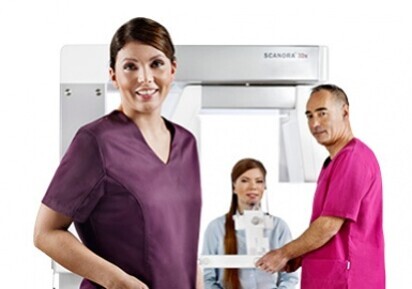
SCANORA® 3Dx
The in-office large field-of-view Cone Beam CT system for Head & Neck imaging
SOREDEX, the imaging system provider from Finland, has introduced a new in-office large field-of-view Cone Beam CT system SCANORA® 3Dx for Head & Neck imaging. The system is intended for wide application areas starting from e.g. single dental implant planning with small FOV up to whole skull imaging with extra large FOV. The system is ideal for ENT (Ear, Nose, Throat), dentomaxillofacial and cranial examinations in imaging centers, ENT offices, total care oral and maxillofacial clinics and hospitals.
The SCANORA® 3Dx is a member of the SCANORA® CBCT product family. Compared to its predecessors, the SCANORA® 3Dx has a larger flat panel detector that enables now a wider range of imaging fields-of-view (FOV) to be used. The same smooth workflow with clear control screen and motorized patient positioning movements characterize this new accuracy instrument. The optional dental panoramic sensor is available as before.
In the unit there are now eight user selectable FOVs available. All the FOVs have their typical applications. The smallest cylindrical FOV 50 x 50 mm with the highest resolution of 0.1 mm voxel (volume element) size is intended for localized problems, such as detailed imaging of single tooth endodontic structures or ossicular chain of the inner ear. Several medium size FOVs are available for imaging for instance both temporal bones in one volume. The most suitable FOV for sinus and ENT imaging is the 140 x 165 mm (HxD) with 0.2 mm voxel size. The largest FOV 240 x 165 mm (HxD) with 0.5 mm voxel size is intended for whole skull examinations, for instance follow-up of facial surgery operations. The voxel volume is isotropic, which ensures that measurements in any direction are accurate.
The SCANORA® 3Dx takes the advantage of the latest imaging technology. The 3D detector is a large amorphous Silicon flat panel for acquiring high resolution projection images. The reconstruction method SARA (SOREDEX Advanced Reconstruction Algorithm) produces 3D volumes out of these projection images.
Accurate patient positioning is achieved with more developed laser lights and scout programs. The seated patient platform ensures perfect stabilization. The FOV can be freely located in any region of interest in the skull area, which makes the system suitable to multiple imaging tasks.
Thanks to wide adjustment ranges of parameters the overall radiation dose for specific diagnostic indications can be optimized by selecting the smallest FOV for each task and adjusting the mA and resolution accordingly.
Contact:
SOREDEX
Nahkelantie 160
04300 Tuusula
Finland


 Austria / Österreich
 Bosnia and Herzegovina / Босна и Херцеговина
 Bulgaria / България
 Croatia / Hrvatska
 Czech Republic & Slovakia / Česká republika & Slovensko
 France / France
 Germany / Deutschland
 Greece / ΕΛΛΑΔΑ
 Hungary / Hungary
 Italy / Italia
 Netherlands / Nederland
 Nordic / Nordic
 Poland / Polska
 Portugal / Portugal
 Romania & Moldova / România & Moldova
 Slovenia / Slovenija
 Serbia & Montenegro / Србија и Црна Гора
 Spain / España
 Switzerland / Schweiz
 Turkey / Türkiye
 UK & Ireland / UK & Ireland
 Brazil / Brasil
 Canada / Canada
 Latin America / Latinoamérica
 USA / USA
 China / 中国
 India / भारत गणराज्य
 Pakistan / Pākistān
 Vietnam / Việt Nam
 ASEAN / ASEAN
 Israel / מְדִינַת יִשְׂרָאֵל
 Algeria, Morocco & Tunisia / الجزائر والمغرب وتونس
 Middle East / Middle East


