Search Dental Tribune

Effective strategies for managing late implant failure and peri-implantitis

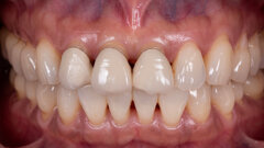
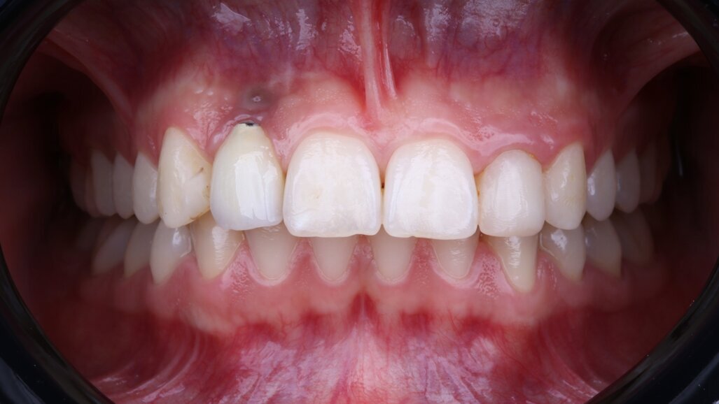
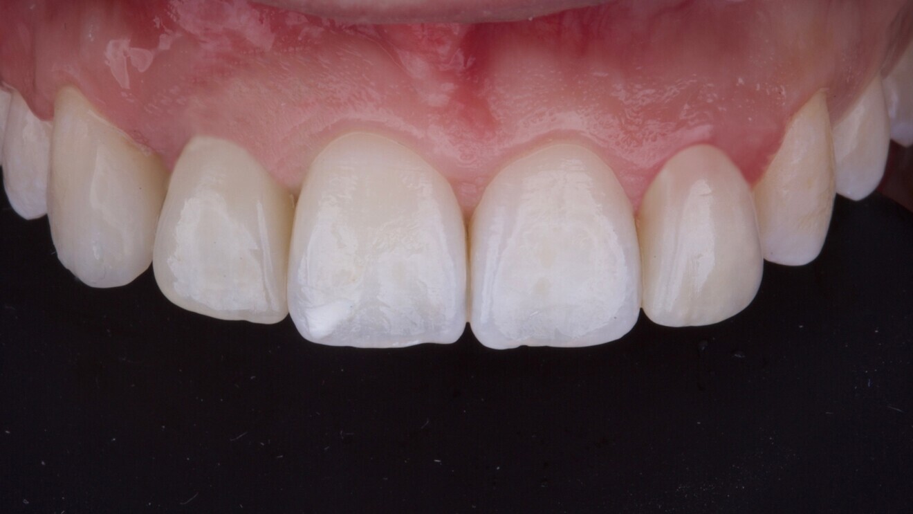
Fig. 1: Peri-implant mucosal inflammation and umbrella effect from bone loss and titanium show-through. (All images: Dr Marco Tallarico)

Late implant failure, particularly in the aesthetic zone, presents significant clinical and biological challenges. This case report describes a digitally guided, biologically sound treatment pathway for managing a failed implant due to peri-implantitis and malposition in a 26-year-old female patient.

The implant was removed using a reverse torque technique, and this was followed by vertical guided bone regeneration using autogenous and xenogeneic grafts and then by soft-tissue augmentation and reimplantation with an implant with a hydrophilic, sand-blasted, acid-etched surface. Digital workflows supported every phase from planning to definitive restoration. One year postoperatively, the implant showed stable osseointegration and optimal soft-tissue architecture, and the patient was very satisfied. This case underscores the importance of peri-implantitis prevention, prosthetically driven positioning and timely intervention in modern implantology.

Dental implants are a cornerstone of modern oral rehabilitation, offering long-term stability and aesthetic outcomes in both partial-arch and full-arch cases. Despite high implant survival rates, the increasing prevalence of implant-related complications—including implant fracture, peri-implant mucositis and peri-implantitis—has highlighted the need for advanced prevention and retreatment protocols.1 Peri-implantitis, in particular, is a multifactorial disease influenced by a range of risk factors, such as plaque accumulation, implant design, prosthetic misfit, occlusal overload and patient-related conditions like smoking or systemic disease.2 Left untreated, peri-implantitis can lead to late implant failure, characterised by progressive bone loss, infection, and aesthetic or functional compromise.

In cases of advanced bone loss or high aesthetic demands, implant removal and replacement often become necessary. While this approach allows for a new start, it presents considerable challenges: the loss of peri-implant bone and soft tissue frequently necessitates advanced reconstructive strategies. Guided bone regeneration (GBR), combined with careful prosthetic planning, is crucial to re-establishing a suitable foundation for future implant placement.3 In this context, digitally assisted workflows—incorporating CBCT, intra-oral scanning and computer-guided surgical protocols—can greatly enhance the precision and predictability of reimplantation procedures.3

Additionally, malpositioning or poor planning of implants is a major contributing factor to long-term biological and prosthetic failure, especially in the aesthetic zone. Even slight deviations from ideal positioning can result in biomechanical overload, prosthetic compromise and eventual tissue breakdown.4 Early diagnosis and timely correction, often through implant removal and site regeneration, are vital for optimal retreatment outcomes.

Recent advances in implant surface technology can improve clinical outcomes in reimplantation cases. Hydrophilic surfaces have demonstrated enhanced wettability and early cellular interaction, promoting faster healing and supporting early osseointegration. In particular, implants featuring sand-blasted, acid-etched surfaces modified with pH-buffering agents—such as the Super Osseointegration (SOI) surface—have shown promising results in enhancing early stability and bone response under early loading conditions.5 These innovations are particularly valuable in cases of compromised bone or when immediate or early loading protocols are indicated after regeneration.

Ultimately, successful management of late implant failure requires an individualised, multidisciplinary approach—one that integrates digital planning, advanced regenerative procedures and biomaterial innovations. The goal is not only to recover lost tissue and restore function, but also to meet the aesthetic expectations of patients through precise, biologically driven protocols.

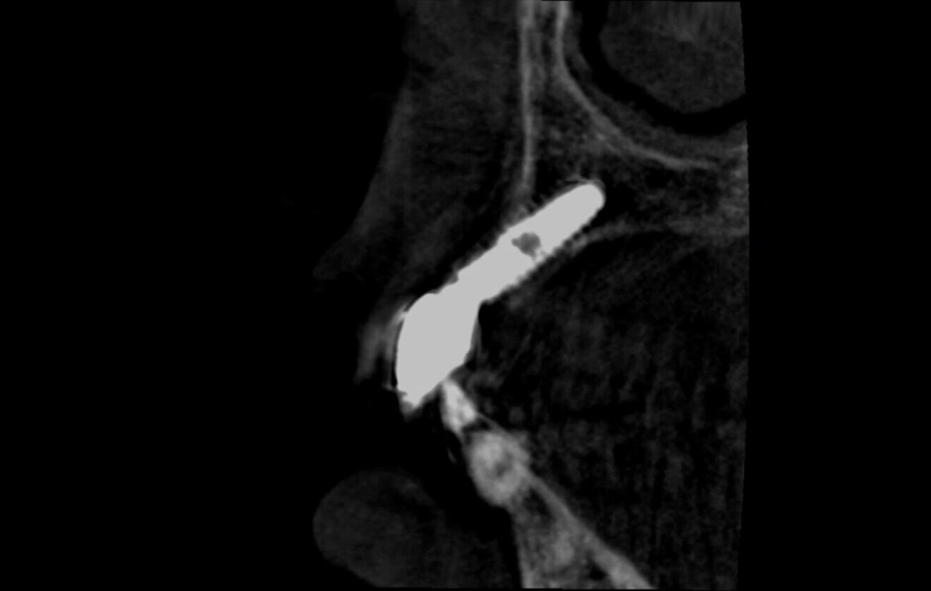
Fig. 2: CBCT evidence of labial implant malposition and bone loss.

Fig. 2: CBCT evidence of labial implant malposition and bone loss.

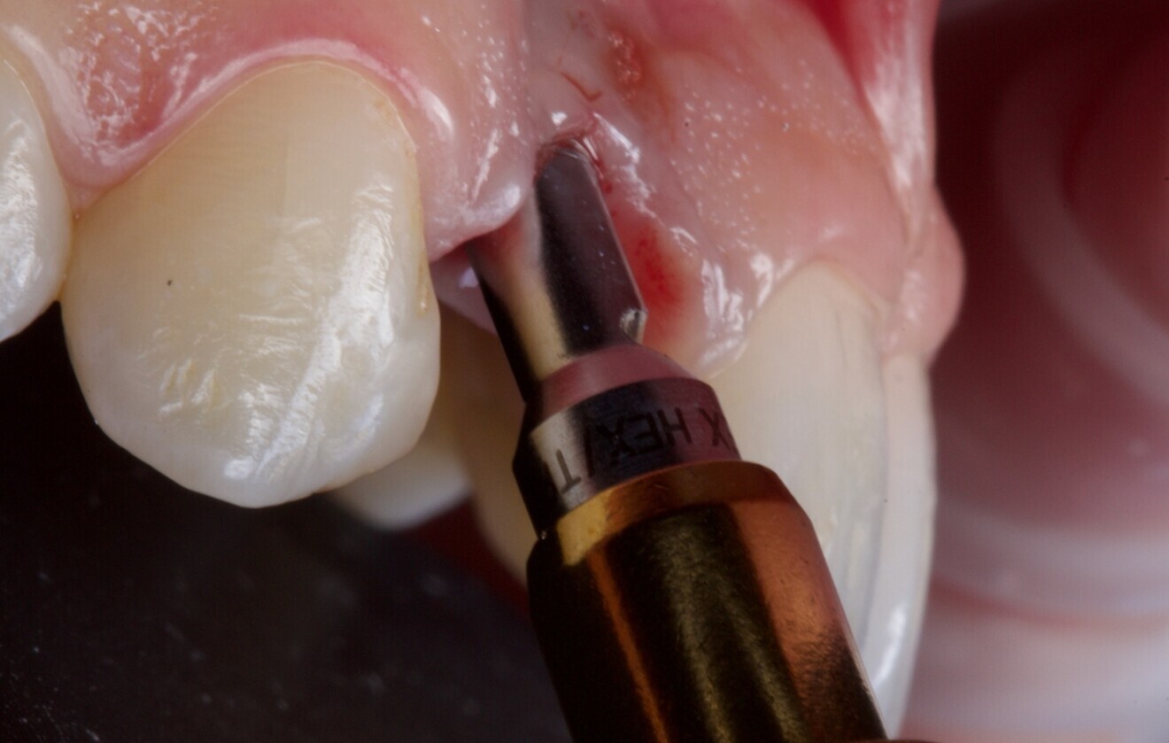
Fig. 3: Atraumatic implant removal using reverse technique.

Fig. 3: Atraumatic implant removal using reverse technique.

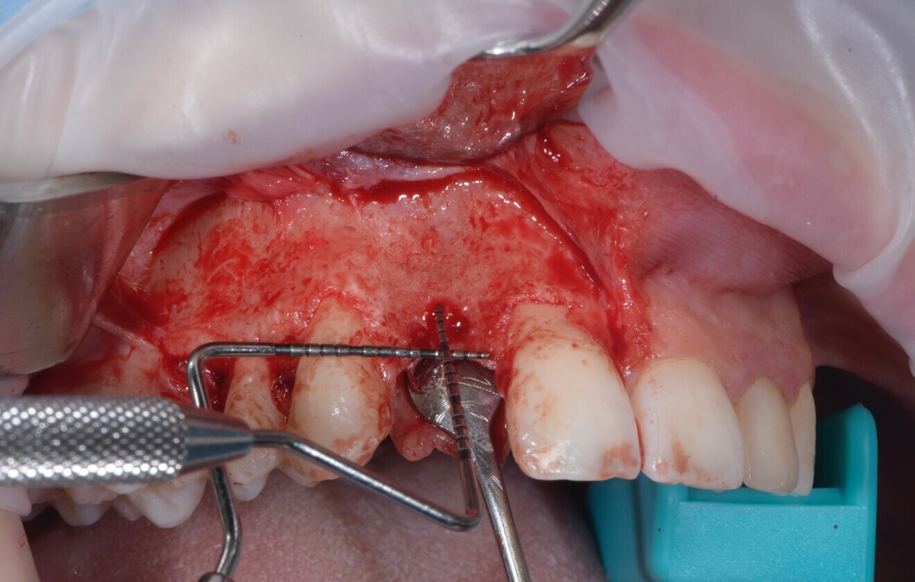
Fig. 4: Vertical guided bone regeneration procedure carried out after an eight-week healing period by experienced operators.

Fig. 4: Vertical guided bone regeneration procedure carried out after an eight-week healing period by experienced operators.

Case summary

A 26-year-old partially edentulous female patient was referred to our clinic with an aesthetic concern in the region of the maxillary right lateral incisor. Clinical examination revealed an osseointegrated but malpositioned implant. The peri-implant soft tissue appeared thin and inflamed. Additionally, a dark-greyish discoloration was visible through the gingiva—referred to as the umbrella effect—caused by the loss of peri-implant bone and show-through of the titanium implant (Fig. 1).

Periapical radiographs showed bone contact on the mesial and distal aspects of the implant. However, clinical probing and CBCT revealed labial and palatal bone loss, consistent with a labially malpositioned implant (Fig. 2).

The patient reported congenital agenesis of the maxillary right lateral incisor and placement of the implant several years prior. After a comprehensive discussion of the treatment options, the patient consented to implant removal and future replacement after bone regeneration. This decision was based on aesthetic concerns and the high risk of further bone loss during medium- to long-term follow-up. The patient was healthy and a non-smoker.

At the initial visit, digital impressions were obtained using the Medit i700 scanner. Periapical radiographs and standardised intra-oral and extra-oral photographs were also acquired. A virtual diagnostic wax-up was generated to guide treatment planning. On the day of surgery, local anaesthesia was administered, and the implant was atraumatically removed using the reverse torque explantation technique. The surgical site was debrided and cleaned, and Type I collagen was applied to the socket (Fig. 3).

A provisional Maryland bridge was bonded to restore the edentulous area aesthetically. After an eight-week healing period, a vertical GBR procedure was performed by two experienced clinicians (MT and SMM). Antibiotic prophylaxis was administered (amoxicillin 2 g 1 hour preoperatively, followed by 1 g twice daily for eight days). The patient also rinsed with 0.2% chlorhexidine for 1 minute before surgery, and the surgical site was isolated with a sterile drape.

Anaesthesia was delivered using 4% articaine with 1:100,000 adrenaline (Ubistesin, 3M ESPE). A crestal incision through the keratinised mucosa was made using a No.15c blade, and a full-thickness flap was elevated. Vertical releasing incisions were placed two teeth away, both mesially and distally (Fig. 4).

The recipient site was debrided, and autogenous cortical bone was harvested from the ipsilateral mandibular ramus (external oblique ridge) using a bone scraper (MICROSS, META). A resorbable collagen membrane (OssMem Hard, Osstem Implant) was secured on the palatal aspect of the defect. A one-to-one mixture of autogenous bone and anorganic bovine bone (A-Oss; particle size: 0.25–1.00 mm; 0.5 g in total; Osstem Implant) was packed into the defect. The membrane was then stabilised with two additional fixation screws.

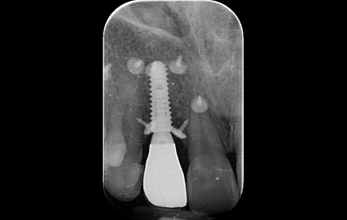
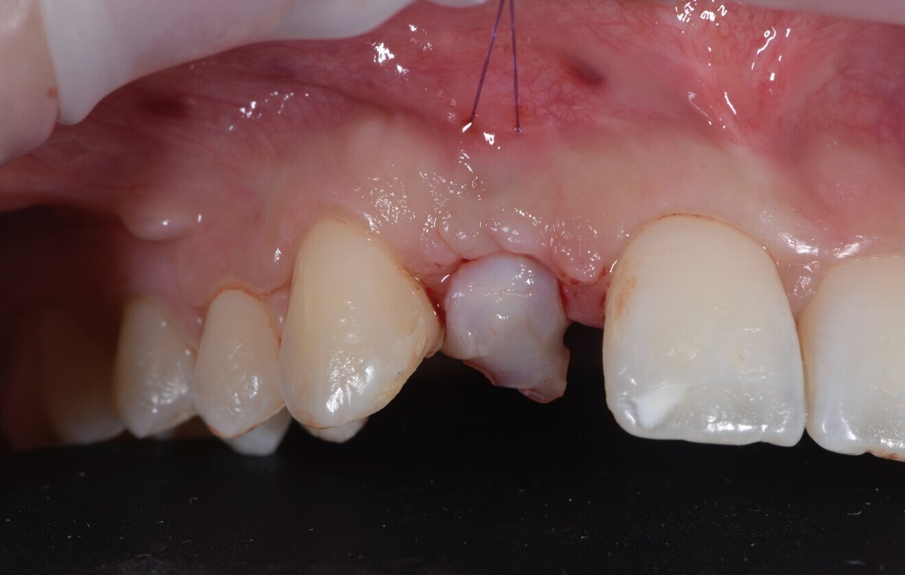
After eight months of uneventful healing, a CBCT scan (6 × 8 cm field of view, 90 kVp, ~ 7 mA) was performed. A prosthetically guided surgical guide was designed to ensure optimal implant positioning. Under local anaesthesia, a flap without vertical releasing incisions was raised. A new implant (TSIII SOI, 3.5 × 11.5 mm; Osstem Implant) was placed using a fully guided protocol (Fig. 5). After implant placement, a connective tissue graft was harvested from the palatal area (first premolar to first molar region) and sutured to thicken the peri-implant mucosa (Fig. 6). The patient was provided with detailed postoperative care instructions and medication.

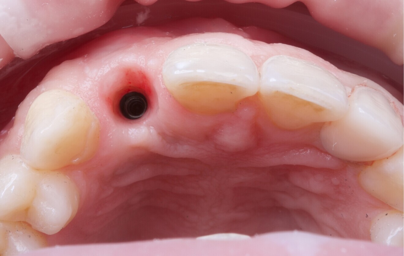
After four months of healing, a minimally invasive uncovering procedure was performed, and a digital impression was taken. Two weeks later, a screw-retained provisional restoration was delivered to contour the peri-implant soft tissue (Figs. 7 & 8).

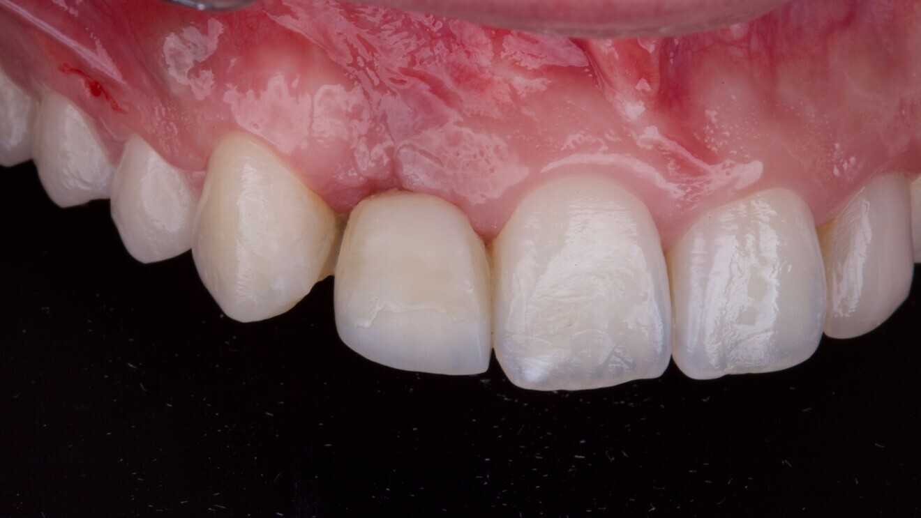
After approximately three months, a final impression was captured, and a definitive porcelain-veneered zirconia crown was fabricated and cemented over a titanium hybrid abutment (Fig. 9). The occlusion was carefully adjusted, and the patient was enrolled in a structured maintenance programme with four-month recall intervals. At the one-year follow-up, the implant demonstrated excellent clinical and radiographic outcomes, showing stable soft tissue and no signs of inflammation or bone loss. The patient reported full satisfaction with the aesthetic and functional results.

Fig. 5: Post-op intra-oral radiograph after implant placement and vertical guided bone regeneration.

Fig. 5: Post-op intra-oral radiograph after implant placement and vertical guided bone regeneration.

Fig. 6: Connective tissue graft from the palate sutured to thicken the peri-implant mucosa after implant placement.

Fig. 6: Connective tissue graft from the palate sutured to thicken the peri-implant mucosa after implant placement.

Fig. 7: Tissue healing after implant placement and connective tissue grafting.

Fig. 7: Tissue healing after implant placement and connective tissue grafting.

Fig. 8: Provisional screw-retained restoration placed to shape the peri-implant soft tissue.

Fig. 8: Provisional screw-retained restoration placed to shape the peri-implant soft tissue.

Fig. 9: Definitive prosthesis delivery: porcelain-veneered zirconia crown cemented on to a titanium link abutment.

Fig. 9: Definitive prosthesis delivery: porcelain-veneered zirconia crown cemented on to a titanium link abutment.

Discussion

This case highlights the multifactorial nature of managing late implant failure, particularly in the aesthetic zone, and underscores the individualised, biologically and prosthetically guided intervention. As implant dentistry matures, clinicians are increasingly confronted with failing implants placed years earlier, often under suboptimal conditions. Late complications such as peri-implantitis and aesthetic compromise are now common, reinforcing the need for comprehensive diagnostic, surgical and restorative planning to prevent risks such as malposition.1

The first critical clinical decision in managing a failed implant is whether to attempt salvage or proceed with removal. This choice must be guided by a combination of scientific evidence, clinician experience and patient-specific factors—including bone loss, soft-tissue status, aesthetic expectations and long-term prognosis. The consensus classification of peri-implant disease emphasises staging and grading to help determine disease severity and appropriate intervention.2 In this case, the implant presented with progressive labial and palatal bone loss and thin soft tissue—both unfavourable prognostic indicators. According to recent consensus, implants showing progressive or circumferential bone loss, especially in aesthetic regions, should be removed promptly to prevent further hard- and soft-tissue compromise.3,4

Once removal was indicated, the use of the reverse torque explantation technique allowed for a minimally invasive and bone-preserving approach. This conservative method has been shown to minimise additional trauma and maintain the integrity of the recipient site, thus supporting future regenerative procedures.5

The cornerstone of successful reimplantation is prosthetically driven implant placement, ensuring ideal 3D positioning relative to the definitive restoration. In this case, initial malpositioning had led to biological and aesthetic failure, illustrating how even minor deviations can cause long-term complications. Proper implant positioning not only facilitates optimal load distribution and soft-tissue management but also supports aesthetic harmony.6 Achieving this requires early digital planning, CBCT-based evaluation and virtual wax-ups to visualise the ideal outcome and design a surgical approach to attain it.

To reconstruct the lost alveolar ridge, vertical GBR was performed. Vertical bone defects remain one of the most challenging indications in regenerative dentistry owing to limited vascularity and higher risk of complications. However, predictable outcomes can be achieved with a structured protocol involving autogenous cortical bone, xenografts and resorbable membranes stabilised by fixation screws.7 The choice of grafting material and membrane plays a significant role in maintaining space and supporting osteogenesis during the healing period.8

A further innovation in this case was the use of a hydrophilically modified sand-blasted, acid-etched implant surface. Implants with high surface energy and wettability have demonstrated superior early bone response, faster osseointegration and better outcomes in grafted or compromised sites.9,10 These benefits are particularly valuable in regenerated bone, where vascularity and healing dynamics are more delicate than in pristine bone.

Despite the clinical success, this case underscores the importance of prevention as the key strategy in modern implantology. Prevention begins with correct implant placement, thoughtful prosthetic design and individualised maintenance protocols. Poor positioning, inadequate planning or neglected peri-implant maintenance significantly increase the risk of late complications. Long-term success hinges not only on surgical skill and biomaterials but also on the clinician’s ability to apply a preventive philosophy from the outset.11

Conclusion

This case exemplifies a comprehensive, digitally guided and biologically sound approach to managing late implant failure in the aesthetic zone. The sequence of atraumatic removal, vertical GBR, soft-tissue grafting and prosthetically driven reimplantation led to an aesthetically and functionally stable outcome. The use of biomaterials—specifically autogenous bone, a xenograft, a resorbable membrane and an implant with a hydrophilic surface—contributed to predictable healing and long-term success. Most importantly, this case reinforces the primacy of prevention: placing implants in the correct position, supported by digital planning and prosthetic foresight, remains the most effective strategy to reduce future complications. In compromised or failing cases, timely intervention—guided by staging and grading systems—can transform biologically challenging scenarios into predictable restorative opportunities.

Editorial note:

This article was published in implants—international magazine of oral implantology Vol. 26, Issue 3/2025.

The complete list of references can be found here.

Topics:
Tags:
To post a reply please login or register
advertisement
advertisement