Implant treatment has evolved into a reliable modality for the replacement of missing teeth. Although rare, complications may occur, and some uncertainty surrounds the treatment of some of these events, especially when restorations are supported by a combination of natural teeth and implants.[1] Often the fabrication of an entirely new restoration is necessary if one or several of the natural teeth need to be removed. Here, we report two cases in which a natural tooth abutment of a restoration supported by implants and natural teeth fractured. We describe the technique used to replace the fractured tooth with an implant, which allowed the re-use of the existing restoration.
Case 1
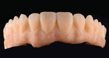
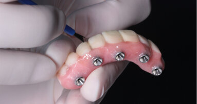
The patient was a 62-year-old male non-smoker in good general health, who was taking no medication and had received implant treatment at the author’s office six years before developing the complications described in this article. The mandible was restored with fixed crowns and implant retained bridges. The maxilla was restored with a removable teeth-implant supported, palatal free bridge (Figs. 1a–d) using double crowns as attachments, as previously described.[2-4]
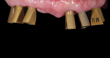
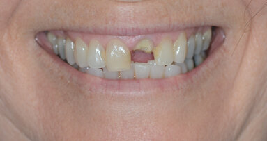
No implants were placed in regio #16 and #26, since the patient decided against performing sinus lift procedures and the remaining bone height was inadequate to allow implant placement. Furthermore, the patient did not agree to extraction of teeth #13 and #23. Therefore, the final restoration had to be supported by four implants (#14, #11, #21, #24; 4.1×10 mm, RN, Straumann, Basel, Switzerland) and two natural teeth (#13, #23) with cantilevers in the areas #15–16 and #25–26 (Figs. 2a–d). Customised implant abutments (torqued to 35 Ncm) and gold copings placed on natural teeth #13 and #23 served as primary telescopes (Fig. 2c). Electroformed pure gold copings with a thickness of 0.25 mm (AGC Galvanogold, Wieland, Pforzheim, Germany), fixated in the superstructure with a self-curing copolymer cement (AGC Cem, Wieland, Pforzheim, Germany), as previously described were used as secondary telescopes.[5,6] The metal framework was milled from a titan 5 alloy (ZENOTEC Ti Disc; Wieland) and covered with microceramic composite (Ceramage, SHOFU, Ratingen, Germany). The patient was put on a three-months maintenance schedule. Six years after implant and prosthetic treatment, the patient reported to the office. Tooth #13 had been fractured in a car accident. He refused any new restoration and insisted on keeping the existing one. Thus, implant placement in position #13 was planned. The fractured tooth #13 was extracted. The maxillary denture was inserted and a bite registration in central occlusion was performed using self-curing acrylic resin (PATTERN RESIN; GC, Alsip, USA). An impression (Impregum; 3M ESPE, Neuss, Germany) of the maxilla was taken with the denture in place. The denture was removed from the patient’s mouth together with the impression (Fig. 3a). This allowed for the fabrication of a cast with an exact duplication of the abutments (Fig. 3b).
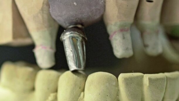
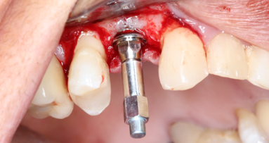
The casts were placed in an articulator using the denture as a guide to achieve correct occlusion (Fig. 4a). A temporary fixed partial denture (from #14 to #24 with #15 and #25 candilevers) from coloured polymethyl methacrylate (PMMA; Zenotec; Wieland, Pforzheim, Germany) was milled based on a scan of the maxilla cast, and was adhered on the abutments using provisional cement (TempBond, Kerr Co., Orange, USA; Fig. 4b). In addition, a surgical stent fitting onto the abutments was milled from clear PMMA (Zenotec; Wieland, Pforzheim, Germany; Fig. 5a). The planned axis of implant #13 was determined using a dental parallelometer (Fig. 5b), and a drill sleeve was placed into the surgical stent (Fig. 5c). An implant (4.5×10 mm, SB line; Dentegris, Duisburg, Germany) was inserted with a torque of 35 Ncm using a two-phase protocol (Fig. 6). Three months after implant placement, an impression post was positioned on the implant and the surgical stent was placed in the patient’s mouth after the drill sleeve had been removed. The impression post was attached to the surgical stent using modeling resin (PATTERN RESIN, GC, Alsip, USA; Fig. 7). After this, the implant analog was attached to the impression post (Fig. 8a) and fixed in the cast using acrylic resin (Fig. 8b). A customised abutment fitting crown #13 was fabricated (Figs. 9a and b), positioned on the implant #13 and torqued to 35 Ncm (Figs. 10a–c). Subsequently, the denture was inserted (Figs. 11a and b).
embedImagecenter("Imagecenter_1_2457",2457, "large");
Case 2
The patient (male, 61 years old and in very good general health) had a foul-mouth periodontal-implant and prosthodontic rehabilitation in 1998. After combined periodontal and implant treatment the mandible was restored with single-fix crowns retained on natural teeth and implants (Fig. 12). The maxilla was restored (in the same way described above for the first case with a removable palatal free metalceramic bridge using double crowns, e.g. telescopic crowns, as attachments, retained on seven natural teeth (#14,13–23) and three implants (#13, 24, 25; RN, 10 x 4.1 mm, Straumann, Basel, Switzerland). Because the patient did not consent to a sinus augmentation, no implants were placed in regio #16 and #26. Tooth #14 was treated endodontically and was used as the last abutment (Fig. 12).
Thirteen years after prosthetic rehabilitation, the patient reported to the office with a root fracture in tooth #15. The tooth was extracted, the secondary telescopic crown in regio #15 was removed, and the supraconstruction was temporarily filled with a photocured, highly elastic temporary material (Fermit, Ivoclar Vivadent, Ellwangen, Germany). The patient again refused a sinus lift and therefore the immediate implant placement regio #15 was scheduled. The axis of the tooth #15, the fabrication of the transfer key and the implant placement, were performed as pre-viously described in case 1. A short implant (Endopore 4.1 × 9 mm, Sybron Implant Solutions, Bremen, Germany) was inserted into area #15 (Fig. 13). Four months after implant placement, impressions were taken and a customised gold implant abutment and new secondary telescopic crown were fabricated and integrated into the same position as tooth #15 (Figs.14 and 15). During a healing period as well as after the integration of the new abutment and the new secondary telescope in the bridge, the patient further used his telescopic maxillary restoration (Figs. 16a–d).
Discussion
The use of natural teeth and implants to support dentures incurs risks that may lead to loss of an abutment and, subsequently, the whole restoration. Recent reports have demonstrated a high long-term success rate of removable restorations supported by natural teeth and implants when double crowns, e.g. telescopic crowns, are used as attachments.[7-9] However, the use of the combination, e.g. connection, of natural teeth and implants to support fixed dentures is not advisable due to the higher risk of complications.[1,10] Cause for the loss of the abutment in case 1 was trauma from a car accident and not mechanical failure or periodontal infection or bone defects. Nevertheless, the resulting complications are similar to the ones described in the literature for cases where the above-mentioned causes lead to loss of an implant.[1]
In case 2, the natural abutment was lost due to mechanical reasons 13 years after loading. This kind of complications, e.g. fractures, have been reported in the long-term maintenance of fixed or telescopic reconstructions, when endodontically treated teeth were used as abutments.[11,12] The complication discussed above could be avoided if the endodontically treated last natural tooth abutment #15 was extracted and replaced by an implant.
In cases of full-arch restorations retained on both implants and natural teeth, when a fracture of a natural abutment occurs, removal of the restoration is often necessary, regardless of the type of restoration (removable or fixed). This can cause not only conflicts between patient and dentist, but also high financial and technical efforts. The technique described above allows the successful replacement of the failed abutment by an implant, enabling the continuous use of the existing restoration.
Editorial note: A list of references is available from the publisher. This article was published in CAD/CAM international magazine of digital dentistry No. 02/2016.
A 72-year-old patient in good general health presented with partial edentulism in the maxillary posterior region. The main goal was to achieve a rapid fixed...
Dental technicians have so many different restorative materials and design and finishing concepts available to them that it can seem difficult to select the...
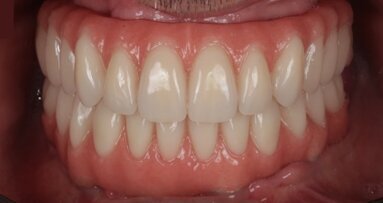
There is increased interest in the long-term clinical outcomes and quality of life of patients treated with full-arch implant-supported dental prostheses ...
A 38-year-old female patient presented to our dental practice complaining of discomfort in the maxillary left area. The clinical examination revealed a ...
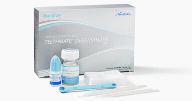
Kuraray Noritake Dental’s TEETHMATE DESENSITIZER is designed to provide an immediate and long-lasting solution for dentine sensitivity. Owing to a ...
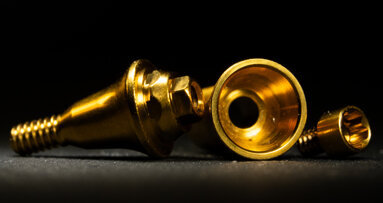
In a free upcoming webinar on 9 June, Dr Stavros Pelekanos will explain the importance of implant abutment design for implant dentistry and discuss the ...
Many factors can be related to apical root resorption and rounding, among them orthodontic movement and occlusal trauma. In severe cases, the tooth can even...
IOANNINA, Greece: Numerous finishing techniques are available for zirconia-based screw-retained implant-supported prostheses; however, most of them are ...
BERLIN, Germany: The number of implants placed is constantly increasing, and so are the possibilities of different prosthetic solutions. Researchers from ...
The long-term clinical success of dental implants is dependent upon osseointegration, which is defined as a direct functional and structural connection ...
Live webinar
Tue. 3 March 2026
11:00 am EST (New York)
Dr. Omar Lugo Cirujano Maxilofacial
Live webinar
Tue. 3 March 2026
8:00 pm EST (New York)
Dr. Vasiliki Maseli DDS, MS, EdM
Live webinar
Wed. 4 March 2026
12:00 pm EST (New York)
Munther Sulieman LDS RCS (Eng) BDS (Lond) MSc PhD
Live webinar
Wed. 4 March 2026
1:00 pm EST (New York)
Live webinar
Fri. 6 March 2026
3:00 am EST (New York)
Live webinar
Tue. 10 March 2026
4:00 am EST (New York)
Assoc. Prof. Aaron Davis, Prof. Sarah Baker
Live webinar
Tue. 10 March 2026
8:00 pm EST (New York)
Dr. Vasiliki Maseli DDS, MS, EdM


 Austria / Österreich
 Bosnia and Herzegovina / Босна и Херцеговина
 Bulgaria / България
 Croatia / Hrvatska
 Czech Republic & Slovakia / Česká republika & Slovensko
 France / France
 Germany / Deutschland
 Greece / ΕΛΛΑΔΑ
 Hungary / Hungary
 Italy / Italia
 Netherlands / Nederland
 Nordic / Nordic
 Poland / Polska
 Portugal / Portugal
 Romania & Moldova / România & Moldova
 Slovenia / Slovenija
 Serbia & Montenegro / Србија и Црна Гора
 Spain / España
 Switzerland / Schweiz
 Turkey / Türkiye
 UK & Ireland / UK & Ireland
 Brazil / Brasil
 Canada / Canada
 Latin America / Latinoamérica
 USA / USA
 China / 中国
 India / भारत गणराज्य
 Pakistan / Pākistān
 Vietnam / Việt Nam
 ASEAN / ASEAN
 Israel / מְדִינַת יִשְׂרָאֵל
 Algeria, Morocco & Tunisia / الجزائر والمغرب وتونس
 Middle East / Middle East








To post a reply please login or register