Search Dental Tribune

Virtual live conference of solidarity—Osstem Meeting Online drives digital transition

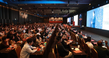
The Osstem Meeting Online is a series of interactive online symposia, which will take place on 12 May and 4, 18 and 27 June. (Image: Osstem Implant)
Osstem Implant

Osstem Implant

Wed. 29. April 2020

save

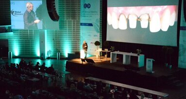
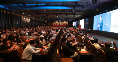
SEOUL, South Korea: On 21 April, Osstem Implant held the first session of its virtual conference, the Osstem Meeting Online, which achieved over 95,000 total live cumulative views from more than 60 countries within 2.5 hours. This series of live-streamed symposia, which create a virtual conference experience for dental professionals around the globe, will culminate in a session on 27 June that will feature a socially sustainable fundraising campaign.

The world is holding its breath in the times of the coronavirus. The COVID-19 crisis is having a profound impact on social life and poses acute economic challenges for many industries, and the dental industry is no exception. Many events, including conferences and conventions, in the field are being cancelled or postponed. Osstem Implant too has announced the postponement of its 13th annual global symposium, the 2020 Osstem World Meeting in Istanbul in Turkey, to next year.

Driving a digital transition of dental conferences

At the same time, however, by employing high technology and new technical solutions for digitalisation, Osstem Implant is taking a crucial leap towards the new era of implantology. Since 2018, Osstem Implant has already been providing dental professionals with opportunities to participate in its annual global meeting by offering live-streaming services. Based on its years of experience in digital transformation, Osstem Implant invites dental professionals around the world to an entirely virtual global conference experience this year. The Osstem Meeting Online will run until 27 June 2020, and the entire programme, including nine lectures and five live surgeries, will come alive via its own interactive live-streaming platform.

Hoping that the global situation will get better, we provide the ultimate virtual live conference experience this year, bringing dental professionals of the world closer to innovative ways of living together, Osstem Implant stated.

Knowledge transfer through real-time interaction

Whether they are at entry level and keen to learn or experienced dental professionals seeking a new challenge, attendees will find relevant standard and highly advanced live surgeries and comprehensive seminars by world-renowned speakers. Allowing the maximum knowledge transfer through real-time interaction between speakers and attendees, the first session on 21 April featured two live surgeries. Covering rare cases of implant placement in partially and completely edentulous patients aged over 70, it successfully delivered first-class implant treatment educational content to participants. All attendees had the opportunity to pose real-time questions and engage further with the speakers and other participants after the session.

Towards solidarity in dental implantology

On 27 June, the conference will celebrate its grand finale with a live surgery by Dr Marco Tallarico from Italy, and this will be combined with a socially sustainable fundraising campaign, in which the speaker, Osstem Implant and all attendees will actively participate and engage with one another. The entire amount of the fund collected will be donated to an organisation in Italy for support in overcoming the COVID-19 crisis.

Register now

Next sessions will be held on 12 May and 4, 18 and 27 June. Online registration for the Osstem Meeting Online will remain open until 27 June. More information about the meeting and registration can be found here or via the social media channels of Osstem Implant.


One thought on “Virtual live conference of solidarity—Osstem Meeting Online drives digital transition

  1. Balakrishnan says:

    I am inerested as an osstem user

To post a reply please login or register
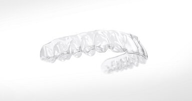
Vertical control using aligners and mini-screws enables predictable, nonsurgical open bite correction through controlled molar intrusion and mandibular auto-rotation, providing stable, long-term clinical results. (Image: edwardolive/Adobe Stock; clinical images: Dr Domingo Martín)

Fri. 24. October 2025

save

Traditionally, anterior open bites have been considered one of the most challenging malocclusions to treat in orthodontics. In fact, the majority of such cases have historically required orthognathic surgery for effective and stable correction.1–3  However, this paradigm shifted significantly with the introduction of skeletal anchorage, which enabled more predictable posterior intrusion and allowed for alteration of the occlusal plane. These biomechanical changes facilitate mandibular auto-rotation, a key mechanism in what is now recognised as vertical control for the nonsurgical closure of open bites.4, 5

Despite these advancements, it is still common to observe clinicians—especially at conferences—attempting to correct open bites primarily through anterior extrusion. Unfortunately, this method has a high tendency for relapse, and recurrence of the open bite is often seen after treatment. In contrast, when the correction is achieved through molar intrusion, the natural masticatory forces tend to support and maintain the closure, making relapse significantly less likely.6, 7

It is important to emphasise that aligners are an ideal appliance for achieving vertical control. The physical thickness of the aligners, combined with occlusal forces during function, can aid in the intrusion of posterior teeth.8, 9

Figs. 1a–f: Pretreatment extra-oral photographs.

Figs. 1a–f: Pretreatment extra-oral photographs.

However, successful outcomes require accurate planning from the start—specifically, the calculation of the necessary amount of intrusion and a clear understanding of the resulting sagittal changes once the vertical discrepancy has been corrected. To achieve this, it is essential to use an articulator mounting (formerly analogue, now virtual) to evaluate the final sagittal relationship after vertical correction.10 Furthermore, it is critical to employ treatment planning software that considers the patient’s actual hinge axis or arc of closure in order to guide mandibular auto-rotation and ensure physiological and stable results.

The purpose of this article is to present a series of open bite cases that were successfully treated at the Martín Goenaga clinic in San Sebastián in Spain using vertical control techniques that combine the aligner with skeletal anchorage through mini-screws.

Case 1: Anterior open bite in a dolichofacial patient

An 18-year-old female patient presented with the chief complaint of an anterior open bite. The patient exhibited a dolichofacial skeletal pattern with a symmetrical facial appearance, a lack of incisor display at rest and an inverted smile line. Her profile was convex due to bimaxillary retrusion, accompanied by an obtuse nasolabial angle (Fig. 1).

Clinical examination revealed a visible anterior open bite, a Class III dental tendency, maxillary constriction, mild crowding in both arches and a deviation of the mandibular midline to the right. The maxillary arch had a triangular shape, whereas the mandibular arch was square in form (Fig. 2).

The panoramic radiograph confirmed the presence of all four third molar germs. Notably, there was advanced root resorption in the maxillary arch, although the overall periodontal health appeared good (Fig. 3). The cephalometric analysis showed a Class II skeletal pattern, a vertical growth tendency and posterior mandibular rotation. The maxillary and mandibular incisors were found to be slightly proclined (Fig. 4).

Basal width measurements on the CBCT scan, using the Penn transverse analysis, revealed a maxillary width of 57.7 mm and a mandibular width of 54.8 mm, yielding a +2.9 mm differential. This indicated that skeletal expansion was not necessary in this case (Fig. 5).

Treatment planning
To provide clear instructions for aligner staging and biomechanics, a virtual treatment objective (VTO) was created. It was determined that 4 mm of posterior intrusion would be required to facilitate mandibular auto-rotation and effectively close the open bite. Accordingly, posterior intrusion was planned in both arches to achieve vertical control and a favourable occlusal outcome (Fig. 6).

Treatment mechanics and biomechanics
The chosen aligner system allowed for simulation of mandibular anterior rotation around the true hinge axis—the arc of closure—which is a critical factor in the successful treatment of anterior open bites.

Using the system’s treatment planning software, we simulated (1) alignment and levelling of both arches (without incisor protrusion), (2) maxillary dentoalveolar expansion, (3) posterior intrusion in both the maxilla and mandible, (4) mandibular midline correction and, ultimately, (5) mandibular auto-rotation. To resolve the mandibular crowding without further proclining the teeth, we incorporated posterior IPR (Fig. 7).

It is important to note that the maximum predictable intrusion with aligners alone is approximately 1 mm in the molar region. Therefore, in cases requiring greater intrusion, it is essential to reinforce vertical movement using skeletal anchorage—in this case, mini-screws and elastics.

At the initial appointment, the attachments were bonded. In the maxilla, mini-screws (1.6 × 10.0 mm) were placed buccally between the first and second premolars and between the first and second molars, and palatally between the first and second premolars. The patient was instructed to wear 5/16 in., 8 oz vertical elastics bilaterally extending from the buccal to the palatal mini-screws over the aligners. In the mandibular arch, mini-screws were inserted buccally between the first and second premolars and between the first and second molars. The patient was instructed to wear elastics from these mini-screws over the aligners to lingually bonded buttons (Fig. 8).

After the 20th aligner, we initiated the reset phase, pausing treatment to rescan the patient and replan the final phase. At this stage, crowding had been resolved, and the open bite had been closed, although additional posterior intrusion was still needed to establish a stable correct overbite. This mid-treatment refinement is crucial to ensure precise fit and continued accuracy of the aligners.

After the reset phase, which involved treatment with 12 aligner pairs, treatment continued with a focus on further maxillary posterior intrusion to fine-tune the vertical correction (Fig. 9). The second round of aligners targeted case finishing with the specific objectives of improving posterior intercuspation, achieving precise midline correction and enhancing sagittal relationships through posterior IPR and the bilateral use of Class III elastics (Fig. 10).

Final outcomes
After 18 months of treatment, including the reset phase and refinements, the patient exhibited significant extra-oral improvements, including a more harmonious smile arc, increased incisor display at rest and notable facial profile enhancement driven by effective mandibular auto-rotation (Fig. 11). Intra-orally, the results included Class I molar and canine relationships, proper alignment and levelling of both arches, an ideal overjet and overbite, and coinciding maxillary and mandibular midlines (Fig. 12).

Beyond the occlusal and aesthetic improvements, vertical control had delivered significant facial changes, including a shortening of the lower facial third, improved upper and lower lip support, and an advancement of the chin by 5 mm (Fig. 13). Importantly, all of these changes were achieved without exacerbating the initial root resorption, as confirmed by the final panoramic radiograph (Fig. 14). The final cephalometric radiograph and tracing confirmed the successful treatment outcome, showing clear evidence of mandibular auto-rotation and a shortened lower facial third, contributing to improved facial proportions (Fig. 15).

Moreover, the final virtual articular mounting demonstrated a precise match between the patient’s maximum intercuspation and true arc of closure. This alignment reflects the achievement of orthopaedic stability—a sine qua non for long-term dental health, functional occlusion and the stability of the treatment results (Fig. 16).

Case 2: Patient with a complex malocclusion and temporomandibular disorder signs and symptoms

A 23-year-old female patient presented with persistent signs and symptoms of temporomandibular disorder (TMD) as her chief complaint. After seeking multiple professional opinions, she came to our clinic for a third evaluation. The patient exhibited a symmetrical facial structure, a bimaxillary retrusive profile and a mildly increased lower facial third. On smiling, dental crowding became apparent (Fig. 17).

Figs. 17a–f: Pretreatment extra-oral photographs.

Figs. 17a–f: Pretreatment extra-oral photographs.

The occlusal analysis revealed a Class II Division 2 malocclusion, along with an anterior open bite tendency, insufficient overjet and overbite, maxillary and mandibular constriction, negative torque in both the posterior segments and maxillary incisors, a pronounced curve of Spee and severe crowding in both arches. The dental midlines were not centred. The mandibular midline deviated to the left, primarily owing to the inclination of the mandibular incisors. Despite having a favourable gingival biotype and adequate attached gingiva, the patient presented with localised gingival recession and abfraction lesions—most likely the result of occlusal instability and functional overload (Fig. 18).

The panoramic radiograph showed prior extraction of all third molars, localised root resorption of several teeth, generally good periodontal health and an atypical condylar morphology, suggestive of temporomandibular joint (TMJ) remodelling or degenerative changes (Fig. 19). The cephalometric radiograph and tracing revealed a mesobrachyfacial skeletal pattern and a tendency towards a Class II skeletal pattern (Fig. 20).

TMJ evaluation
The patient’s main concern of uncertainty in mastication is a classic indicator of occlusal instability. Additional symptoms included nocturnal bruxism, morning muscle fatigue and occasional episodes of mandibular locking.

A virtual articulator mounting using MODJAW revealed a significant discrepancy between maximum intercuspation and centric occlusion (Fig. 21). CBCT imaging of the joints further confirmed joint pathology, showing inferior displacement of the right condyle, flattened articular surfaces on both condyles and a bifid morphology of the left condyle (Fig. 22). Given the presence of TMD symptoms, pronounced discrepancy between maximum intercuspation and centric occlusion, and clinical signs of gingival recession and abfraction lesions—all consistent with joint instability—we initiated therapy with a condylar repositioning appliance.

Splint therapy
We selected a two-piece FACE splint, worn 24 hours a day in the posterior segment during the day and the anterior segment at night (Fig. 23). The splint therapy lasted for four months, during which a stable condylar position was achieved, the patient transitioned to a single arc of closure and full remission of TMD symptoms was obtained. As a result of mandibular posterior rotation, the occlusion changed, presenting with an increased anterior open bite (Fig. 24), an increased overjet (Fig. 25) and an increased Class II dental relationship (Fig. 26).

Treatment planning
A VTO based on the post-splint cephalometric tracing outlined the biomechanical needs for resolving the case and reaching our goals of proclination of the maxillary incisors by 9°, intrusion of the mandibular incisors by 2 mm to level the curve of Spee, and intrusion of the maxillary posterior segments by 3 mm to facilitate closure of the open bite through mandibular auto-rotation. This comprehensive plan was then communicated to the digital aligner planning team for execution (Fig. 27).

Treatment mechanics and biomechanics
In the 3D digital treatment set-up, both maxillary and mandibular arch forms were improved by applying positive torque to the posterior segments, facilitating proper alignment and levelling. Specific planned movements included proclination of the maxillary incisors, and retraction and intrusion of the mandibular incisors. IPR in the mandibular arch was planned in order to relieve crowding without further proclination.

Since the required intrusion of the maxillary posterior teeth to enable mandibular auto-rotation exceeded 1 mm, interradicular mini-screws were indicated to enhance force application and ensure controlled vertical movement (Fig. 28). At the first clinical appointment, the attachments were bonded according to the digital set-up. A total of six mini-screws were placed in the maxilla: four buccally between the first and second premolars and two palatally between the first molar and second premolar. The patient was instructed to wear 5⁄16 in., 8 oz elastics bilaterally over the aligners and attached to the mini-screws in order to facilitate posterior intrusion (Fig. 29).

The active orthodontic treatment was carried out with 60 maxillary and mandibular aligners, delivered in phases: 30 aligner pairs or the initial phase, 18 aligners for the first refinement and 12 aligner pairs for the second refinement. In the final phase of treatment, 3⁄16 in., 4.5 oz intermaxillary elastics were prescribed to improve the Class II dental relationship and enhance final intercuspation. The total treatment time was 18 months, including periods where the patient wore Essix-type retainers between refinement phases.

Final outcomes
Extra-orally, enhanced smile aesthetics was achieved thanks to the maxillary arch expansion and resolution of crowding. There was also a shortening of the lower facial third, improved chin projection and enhanced lip support (Fig. 30).

Intra-orally, both arches showed improved shape (expanded by approximately 1.5 mm per side), well-aligned and levelled teeth, and resolution of the initial crowding. Class I molar and canine relationships were achieved bilaterally, as well as good final overjet and overbite. The maxillary incisors were proclined (U1–PP from 93° to 106°), and the mandibular incisors were slightly retruded (IMPA from 99.4° to 97.2°). The midlines were centred (Fig. 31).

The post-treatment panoramic radiograph showed good root parallelism and no worsening of the pre-existing root resorption or periodontal condition (Fig. 32). The final cephalometric radiograph and tracing showed that, although the patient retained a Class II skeletal pattern, the values had improved owing to mandibular auto-rotation (ANB from 6.9° after splint therapy to 4.4° at the end of treatment). This auto-rotation had also improved the vertical measurements between the end of splint therapy and the end of treatment: maxillary inclination (SN–OP) had decreased from 18.6° to 16.2°, Ricketts’ facial axis from 92.7° to 91.4° and Jarabak’s sum of angles from 392.0° to 387.6° (Fig. 33).

Finally, and most importantly, orthopaedic stability had been achieved, and there was no discrepancy between maximum intercuspation and centric occlusion (Fig. 34). There was also complete resolution of the TMD symptoms.

Case 3: Post-aligner treatment case with signs and symptoms of TMD

This case involves a 29-year-old female orthodontist who was completing a fellowship at our clinic. During her time with us, she began to suspect that her own symptoms might be related to her occlusion. Although she had previously undergone orthodontic treatment, she was now experiencing TMJ discomfort. After observing the successful treatment of several patients for similar TMJ issues, she approached us for a diagnostic evaluation of her own case.

Figs. 35a–f: Pretreatment extra-oral photographs.

Figs. 35a–f: Pretreatment extra-oral photographs.

She presented with a symmetrical face, insufficient incisor display at rest and increased gingival display in the posterior segments during smiling. Hypertrophy of the masseter muscles was noted (Fig. 35). Class I molar and canine relationships, well-aligned arches and a favourable arch form were observed. The dental midlines were centred. However, there was a tendency towards anterior open bite, absence of both overjet and overbite, and negative torque of the maxillary incisors. Additionally, the patient exhibited signs of dental wear and mandibular tori—both common findings in patients with bruxism or clenching habits (Fig. 36).

The panoramic radiograph revealed the absence of third molars and good root morphology and parallelism (Fig. 37). The cephalometric radiograph and tracing indicated a Class III skeletal pattern tendency, characterised by a short cranial base and a long mandible. The maxillary incisors were retroclined, and the mandibular incisors were proclined (Fig. 38).

TMJ analysis
The patient reported joint and orofacial muscle pain, headaches, neck pain and nocturnal bruxism. CBCT imaging showed small condyles with flattened articular surfaces. The right condyle was slightly posteriorly positioned within the mandibular fossa (Fig. 39). Additionally, a virtual articulator mounting using the MODJAW system revealed that there was a significant discrepancy between maximum intercuspation and centric occlusion and that the first occlusal contact was at tooth #27 (Fig. 40).

Splint therapy
Given these findings, treatment was initiated with a two-piece FACE splint, designed to reposition the condyles and establish a stable occlusal relationship. The splint was worn for five months, during which time a stable condylar position, a single arc of closure and complete remission of TMD symptoms were achieved (Fig. 41). As demonstrated in the initial virtual articulator mounting, splint therapy produced a notable increase in the anterior open bite (Fig. 42).

Treatment mechanics and biomechanics
Orthodontic treatment was planned using a hybrid approach, combining fixed appliances and aligners to efficiently address the malocclusion. Treatment began with the placement of bite raisers on teeth #16, 17, 26 and 27, precisely positioned according to the patient’s arc of closure. These served the key purposes of maintaining the stable condylar position achieved by the splint therapy and of facilitating posterior intrusion to support vertical control.

Tubes and brackets were bonded to teeth #14, 15, 16, 17, 24, 25, 26 and 27 and connected using sectional arch wires. Additionally, buttons were bonded to the palatal surfaces of these same teeth. To support vertical control, eight interradicular mini-screws (1.6 × 10.0 mm) were strategically placed—four per side, buccally and palatally between the first and second premolars and between the first and second molars in the upper arch. C-chains were used to deliver intrusive forces from the screws to the brackets and palatal buttons (Fig. 43).

Aligner therapy was simultaneously begun in the mandibular arch, targeting:
  • arch form refinement;
  • positive torque in the posterior segments;
  • improved transverse coordination;
  • elimination of premature contacts; and
  • support for anterior bite closure (Fig. 44).
This first hybrid phase lasted for only three months, during which time effective maxillary posterior intrusion was achieved. As a result, the anterior open bite rapidly closed via mandibular auto-rotation, highlighting the efficacy of the hybrid technique. Moreover, this approach significantly reduced the number of aligners required and shortened the total treatment time (Fig. 45).

Once the bite had closed, treatment continued with aligners in both arches. The goals at this stage were to apply positive torque to the maxillary incisors and canines and to slightly retract the mandibular incisors, as well as to use IPR to improve the overjet and achieve ideal posterior intercuspation (Fig. 46). A total of ten maxillary and mandibular aligners were planned, followed by an additional refinement phase with another ten aligners per arch for finishing and detailing. The total treatment time, including the initial splint therapy, was 17 months.

Final outcomes
Extra-orally, two aspects are worth highlighting: the significant improvement in the smile, in that the patient no longer showed posterior gingiva, owing to intrusion of the maxillary posterior teeth; and the slimmer facial appearance, thanks to muscle relaxation and reduction in the size of the masseter muscles (Fig. 47). Intra-orally, Class I molar and canine relationships had been achieved, and the anterior open bite had been closed, resulting in proper overjet and overbite. The positive torque applied to the maxillary and mandibular posterior segments had improved the arch form. The maxillary incisors were proclined (U1–PP from 105° pretreatment to 112° post-treatment) and the mandibular incisors retroclined (IMPA from 98° pretreatment to 93° post-treatment; Fig. 48).

The final panoramic radiograph showed good root parallelism and confirmed that all treatment objectives had been met without root resorption or harm to periodontal health (Fig. 49). The final cephalometric radiograph and tracing confirmed the mandibular auto-rotation: a facial axis of 95° pretreatment to 97° post-treatment and an anterior facial height of 117.5 mm pretreatment to 115.9 mm post-treatment (Fig. 50).

Finally, the treatment concluded with orthopaedic stability (maximum intercuspation coincided with centric occlusion), as well as with improved condylar position and morphology. The patient was also completely free of TMD symptoms (Fig. 51).

Conclusion

Vertical control using aligners and mini-screws has proved to be highly effective for closing anterior open bites, producing stable long-term results. The use of an aligner system that incorporates the patient’s arc of closure into its planning software is particularly valuable for accurate planning of mandibular rotation and the precise positioning of the incisors. By utilising the arc of closure during treatment planning, we can avoid unnecessary back-and-forth movements, which typically lead to the use of more aligners, an increased number of refinements and longer treatment duration.

Editorial note:

This article was published in aligners—international magazine of aligner orthodontics vol. 4, issue 1/2025. The list of references can be found here.

Topics:
Tags:
To post a reply please login or register
advertisement
advertisement