From the early 20th century, when Walter Hess and Ernest Zürcher [1] demonstrated root canal anatomy with an unprecedented visual clarity, its complexity has fascinated researchers armed with ever better imaging tools—from blue dyes to CT, from CBCT to confocal microscopy, from clear tooth preparations to micro-CT [2–4], to name just a few. Thanks to rigorous research and discussion, the diverse intricacy of root canal morphology is well understood and accepted today. However, the question of how to best prepare this space to restore homeostasis remains open to debate, which is conducted both in the scientific and, unfortunately, commercial domains. Our task as scholars and clinicians is to investigate which approaches would be practical and applicable to bring teeth and periodontium back to health in accordance with evidence-based endodontics and principles of minimally-invasive dentistry.
As yet another array of new file systems are launched in the market, we seem to share an understanding that files do not have the ability to clean root canal space, only preparing, i.e. shaping it, while it is the irrigation process that provides a level of cleanliness that can, hopefully, create conditions for the body to heal. Thus, given that the shaping is acceptable (i.e. the files used remove the bulk of the pulp and/or infected dentine without blocking the system with debris as well as maintain the original shape of the canal without any micro-crack formation), it is the chemical preparation that is responsible for treating the system in all its complexity.
For a long time, irrigation remained a somewhat mystical part of the process, with a general agreement that a good rinse is necessary, but without a standardised sequence of irrigation. While various tools for irrigation and activation of solutions were studied extensively, the first sequence was suggested only in 2005, [5, 6] and it made clinicians aware that alternating solutions could be as beneficial as the use of negative pressure in order to achieve a clean root canal space and diminish postoperative pain.
Below you will find descriptions and outcomes of several studies that led to a suggested protocol of irrigation that is presented in the conclusion of the present publication.
Investigating irrigation today
The fact that during root canal shaping the system may get blocked by debris led to the question of how to best conduct the chemical preparation so that the dentinal tubules remain open to allow for a better cleaning and, consequently, sealing of the system. Drawing from clinical experience and improved outcomes, Jaramillo et al. have formulated an experimental irrigation sequence based on Sleiman’s 2005 suggestions, and added a negative pressure device to see if it may have added benefits [7].
Scanning electron microscopy used to evaluate the cleanliness of dentinal tubules at three different levels of the canals demonstrated that our experimental sequence—alternating the use of 6 percent NaOCl and 17 percent EDTA with water in between—had shown a significantly better ability to keep the entrances of dentinal tubules open and avoid the blockage of dentinal tubules by the smear layer and debris during the cleaning and shaping procedure compared with the use of 6 percent NaOCl or 17 percent EDTA alone. The results emphasised the importance of the early use of 17 percent EDTA and not only as a final rinse.
This sequence allows us to use the standard endodontic irrigants during chemical root canal preparation and prevents any chemical interaction between them thanks to the use of distilled water at strategic times. Depending on the pH levels and the nature of the solutions, such chemical interactions may have a variety consequences, from brown (and in some instances, carcinogenic) precipitation to dentine modification, potentially affecting general health and/or quality of the dentine inside the root canal system, which, in turn, may influence the longevity of the link between the sealer and the dentine, thus changing the outcome of the root canal treatment in general.
Another finding of the study that echoed positive clinical outcomes related to the use of negative pressure in combination with the experimental irrigation sequence; the irrigation protocol that included both the Sleiman sequence (alternating between sodium hypochlorite, water, and EDTA) and a negative pressure irrigation device was proven to be the most efficient in opening dentinal tubules and maintaining them open. It may be posited that the negative pressure allows for a formation of a temporary partial vacuum force, which first draws the liquids from the access cavity into the root canal system and then suctions them out of the system.
Using the macro- and the micro-cannulas of the negative pressure irrigation unit in, correspondingly, the coronal-middle and apical parts of the root canal system, leads to the creation of a vacuum, or a partial vacuum, to be more specific, inside the root canal space. Though its main role is to attract solutions deeper and deeper into the system and safely remove them from within, the partial vacuum created by the negative pressure has a number of other important benefits as Sleiman-Iandolo testing has shown.
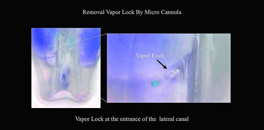
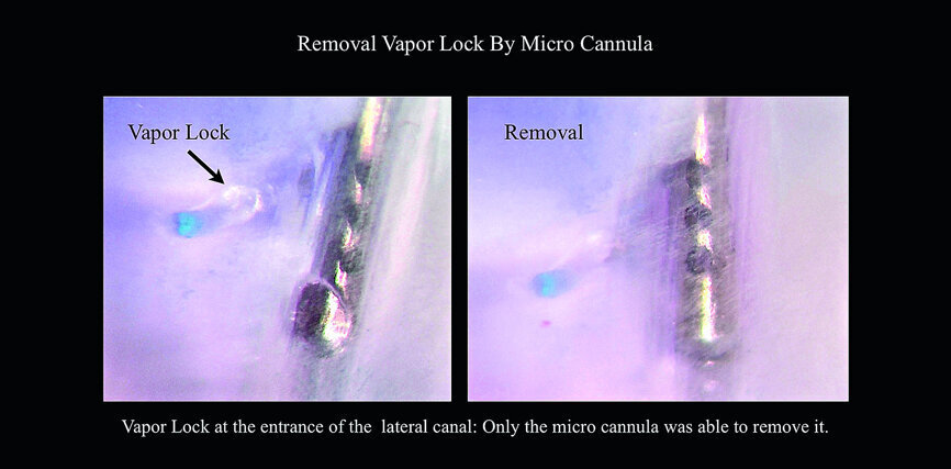
First of all, it can eliminate the airlock (better known in endodontics as vapor lock) inevitably resulting from bubbly chemical reactions between irrigating solutions and the content of the root canal space inside the main canal—mainly in the apical part—as well as inside lateral canals and dentinal tubules and preventing the irrigants from reaching these areas and performing their best (Figs. 1a and b). Secondly, once the airlock is eliminated, the partial vacuum force helps in distributing irrigants into the totality of the root canal system, including the depth of the dentinal tubules. Thirdly, negative pressure irrigation allows for introducing a significantly larger volume of irrigating solutions over a shorter period of time, increasing the efficiency and decreasing the length of the procedure. These unique properties result in a faster and better chemical preparation of the entire internal space.
Fig. 1a: Sleiman-Iandolo testing. Showing a gas bubble locked inside a lateral canal as a result of passive ultrasonic irrigation (PUI).
Fig. 1b: Sleiman-Iandolo testing. Demonstrating the airlock eliminated with the use of the negative pressure irrigation system (NB: other means such as further PUI or use of lateral-vented needle were not helpful in overcoming the vapor lock once produced, the gas bubble remained in place).
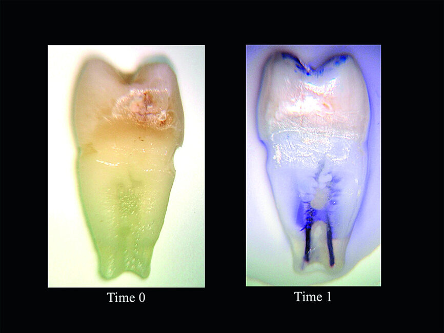
Fig. 2a: Sleiman-Iandolo testing.
Freshly extracted premolar dyed
with methylene blue in a centrifuge.
Note the deep penetration of the dye,
especially in the coronal part.
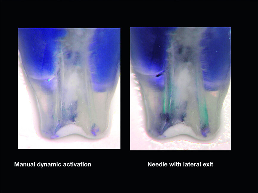
Fig. 2b: Sleiman-Iandolo testing.
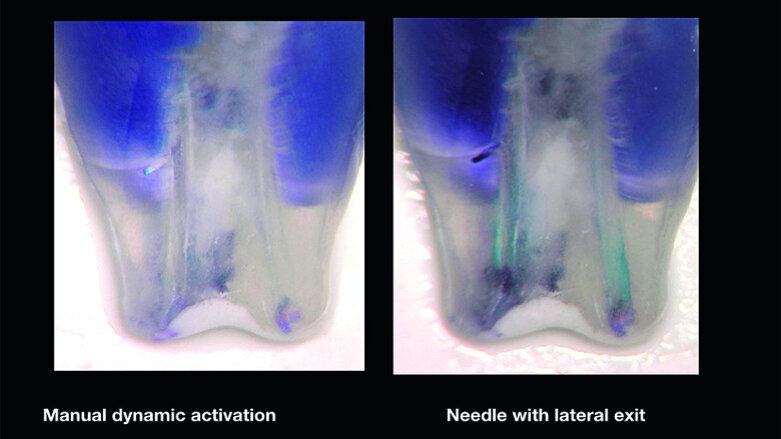
Results of the dye removal with
manual dynamic activation (left) and
side-vented needle (right).
Note the poor results of the cleaning.
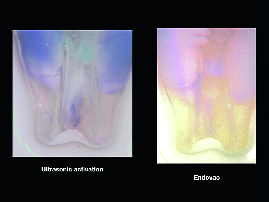
Fig. 2c: Sleiman-Iandolo testing. Results of the dye removal with PUI (left) and a negative pressure irrigation unit used at the intervals suggested as part of the Sleiman sequence (right). Note the extreme cleanliness of the deepest recesses of the dentinal tubules in the image on the right.
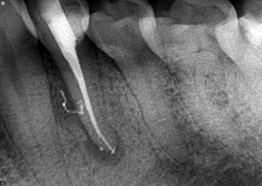
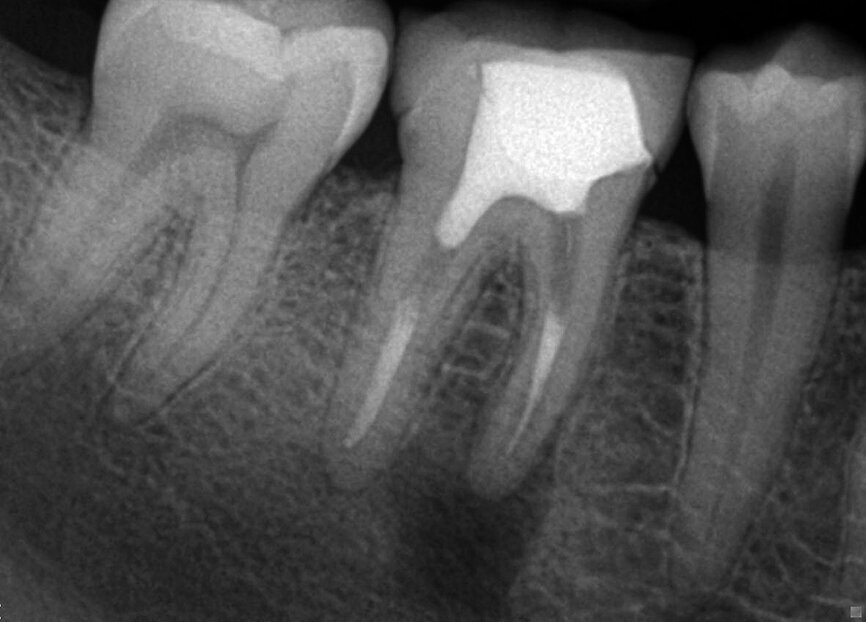
Fig. 3: A necrotic/infected case: Lower premolar of the patient complaining of dull and pulsating pain. The infection was not limited to the apical part, but was also located in the lateral mid-root section. Postoperative X-ray shows a 3-D sealing of the apical area with its anastomosis and of the lateral and accessory canals in mid root.
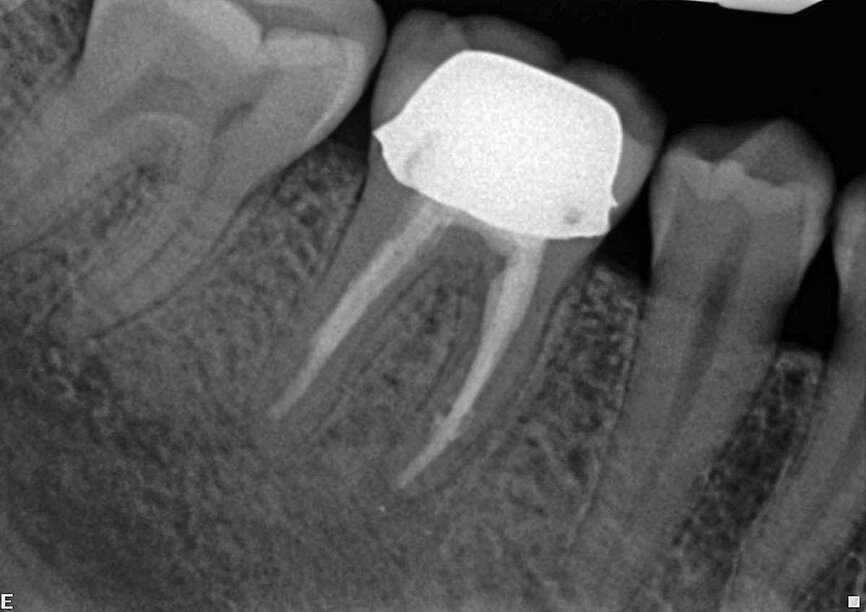
Fig. 4: Complexity of the apical area manifests itself after it was treated chemically, dried, and sealed by way of the warm vertical technique.
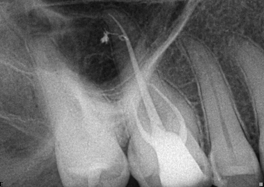
Fig. 5: The case was referred for retreatment due to the failure of the previous root canal therapy. Following the shaping (TF-adaptive) and cleaning (Sleiman protocol), and upon the challenging but successful search for the distal canal, a 3-D obturation was performed, which allows showing the isthmus between the mesial and distal canals as well as a very coronally located lateral canal in the palatal root.
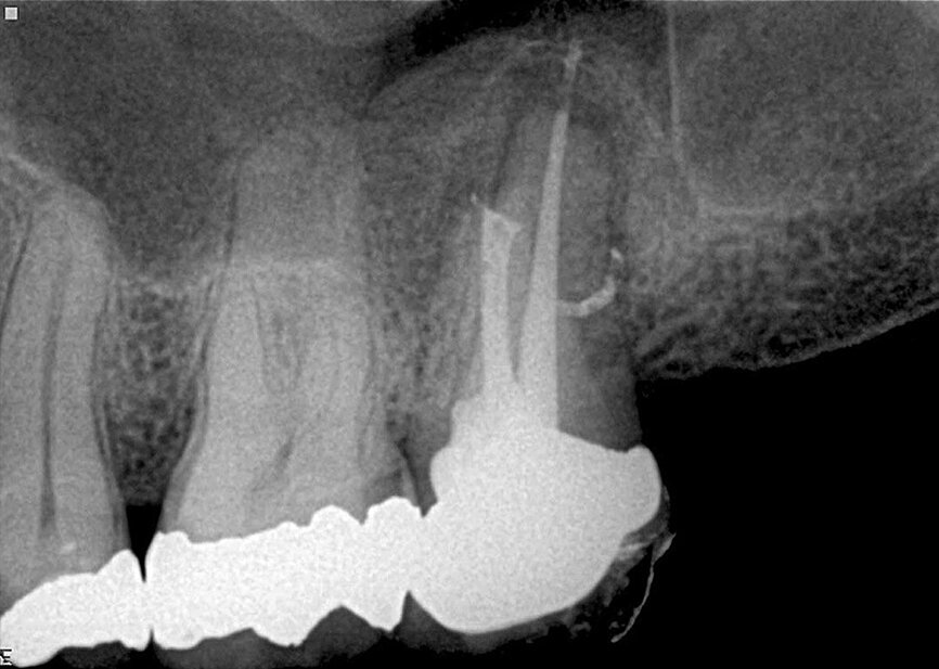
Fig. 6: Vital first upper molar with irreversible pulpitis. Note the extremely long palatal root (approx. 31 mm), nevertheless we managed to seal a beautiful internal loop with a lateral canal, a sign of the proper chemical preparation under partial vacuum and also well dried, allowing the sealer to be compacted inside.
Fig. 7: A failing root canal treatment with apical infection and an internal resorption in the apical area.
Fig. 8: The follow-up confirms a fast healing of both the apical area and the area of the resorption lesion.
Sleiman-Iandolo testing used freshly extracted premolars, removed due to periodontal pathology, impregnated with methylene blue dye in a centrifuge; this resulted in pushing the dye deeply into the dentinal tubules (Fig. 2a). To compare commonly used irrigant delivery techniques, a negative pressure irrigation unit was used (EndoVac) as well as a lateral-vented needle, manual activation of the solution, and passive ultrasonic irrigation in combination with the Sleiman irrigation sequence. EndoVac + Sleiman sequence was shown to be the only approach that allowed for a complete removal of the methylene blue dye from the entire root canal system and dentinal tubules over the total time of 25 minutes, while the other approaches failed to achieve a completely clean system (Figs. 2b & c).
The Sleiman sequence goes beyond using water as an intermediate between the two alternating solutions and as the final irrigant (water cooled to between 2.5°C and 4°C and used for postoperative pain control or in a cryotherapy modality also suggested by Sleiman [8] and investigated by Vera et al. [9])—it also stipulates that when using the macro- or the micro-cannula of the negative pressure irrigation unit for chemical preparation, every five seconds a two-to-three-second pause should be made when no irrigant is added. It is during this pause that the partial vacuum is created by the cannula, which will draw out all the fluids, residues and gases from all the root canal system. Once the system has been drained, the partial vacuum established inside the root canal system in its entirety can attract a fresh portion of irrigant for a faster and cleaner preparation of the root canal system.
Clinical cases
In the images above, we present some of the typical cases demonstrating the cleanliness of the root canal system achieved as shown by the lateral and/or accessory canals visualised upon 3-D warm vertical condensation (Figs. 3–6).
The case of a failing root canal treatment with apical infection and an internal resorption in the apical area was referred to us (Fig. 7). After removing the previous filling, chemical preparation was performed, with the help of the partial vacuum inside the system the chemicals were able to clean the resorption area without an aggressive effect on the periodontal ligament; this has led to a truly three-dimensional obturation. The 4-month follow-up image (Fig. 8) confirms a fast healing of both the apical area and the area of the resorption lesion.
Conclusions
Realising that a 100 percent disinfection of the root canal space remains unattainable, we continue to strive for perfection in our attempts to develop viable clinical protocols that would allow lowering the inflammatory and/or bacterial load so that our patients’ bodies can heal. Based on the supporting research and testing as well as on a history of sustainably high treatment outcomes for both primary endodontic treatment and retreatment of vital and non-vital teeth, we would like to propose our irrigation protocol as a fast, safe, and, most importantly, evidence-based technique of chemical preparation.
The Sleiman irrigation protocol requires 6 percent (or 5.25 percent , if the 6 percent concentration is not available) NaOCl, 17 percent EDTA, distilled water or normal saline. For the best results it is recommended to use a negative pressure irrigation unit to introduce and remove the solutions in order to benefit from the partial vacuum force; however, it must be said that using other introduction techniques in combination with the Sleiman sequence of irrigants will also improve chemical preparation results and lead to a cleaner root canal space.
- Access cavity; manual files to locate orifices; manual files for initial scouting—NaOCl
- H2O
- Machine files for root canal preparation—EDTA
- H2O
- In between machine files—NaOCl
- H2O (cold for cryotherapy)
- Drying the root canal system—EndoVac
The whole irrigation procedure should follow the ‘5 sec introducing solution + 3 sec pause’ guideline to achieve the best effect of the partial vacuum.
Editorial note: A complete list of references is available from the publisher. This article was published in roots - international magazine of endodontology No. 03/2017.
The purpose of preparing of the root canal system is well understood and contemporary techniques involve the use of both hand and rotary instruments used in...
“Faster, higher, stronger”—this motto certainly no longer applies only to the Olympic Games. How can certain tasks be performed even more precisely, ...
After access preparation and location of anatomy, the next challenge facing the endodontic clinician is to select the proper file alloy and sequence for the...
Irrigation is a major step in endodontic treatment. A variety of chemicals are used to achieve what I like to consider the chemical preparation of the ...
Much has changed in endodontics during the last 20 years, except the anatomy, which is still just as complex. We can improve our protocols and techniques, ...
Dr Sergio Rosler is a leading practitioner in the field of endodontics. On Wednesday, 8 April, he will be presenting a webinar that focuses on the ...
To make the wait for ROOTS SUMMIT 2024 a little sweeter, the organisers would like to spotlight some of the speakers for next year’s edition, which will ...
The population is ageing rapidly because of the prolonged life expectancy evident in most industrialised countries in the world. Accordingly, the number of ...
Endodontic treatment can attain success rates of between 85 and 97%.1 Adequate treatment protocols, knowledge and infection control are essential to ...
Treating bone insufficiency is a familiar challenge for all implant practitioners. Such insufficiency can compromise the placement of an implant, its ...
Live webinar
Tue. 24 February 2026
1:00 pm EST (New York)
Prof. Dr. Markus B. Hürzeler
Live webinar
Tue. 24 February 2026
3:00 pm EST (New York)
Prof. Dr. Marcel A. Wainwright DDS, PhD
Live webinar
Wed. 25 February 2026
11:00 am EST (New York)
Prof. Dr. Daniel Edelhoff
Live webinar
Wed. 25 February 2026
1:00 pm EST (New York)
Live webinar
Wed. 25 February 2026
8:00 pm EST (New York)
Live webinar
Tue. 3 March 2026
11:00 am EST (New York)
Dr. Omar Lugo Cirujano Maxilofacial
Live webinar
Tue. 3 March 2026
8:00 pm EST (New York)
Dr. Vasiliki Maseli DDS, MS, EdM


 Austria / Österreich
 Bosnia and Herzegovina / Босна и Херцеговина
 Bulgaria / България
 Croatia / Hrvatska
 Czech Republic & Slovakia / Česká republika & Slovensko
 France / France
 Germany / Deutschland
 Greece / ΕΛΛΑΔΑ
 Hungary / Hungary
 Italy / Italia
 Netherlands / Nederland
 Nordic / Nordic
 Poland / Polska
 Portugal / Portugal
 Romania & Moldova / România & Moldova
 Slovenia / Slovenija
 Serbia & Montenegro / Србија и Црна Гора
 Spain / España
 Switzerland / Schweiz
 Turkey / Türkiye
 UK & Ireland / UK & Ireland
 Brazil / Brasil
 Canada / Canada
 Latin America / Latinoamérica
 USA / USA
 China / 中国
 India / भारत गणराज्य
 Pakistan / Pākistān
 Vietnam / Việt Nam
 ASEAN / ASEAN
 Israel / מְדִינַת יִשְׂרָאֵל
 Algeria, Morocco & Tunisia / الجزائر والمغرب وتونس
 Middle East / Middle East













To post a reply please login or register