Dr Sergio Rosler is a leading practitioner in the field of endodontics. On Wednesday, 8 April, he will be presenting a webinar that focuses on the management of complex endodontic anatomy using NiTi files made by the Chinese company Shenzhen Perfect Medical Instruments. Prior to the online lecture, Rosler spoke to Dental Tribune International about the details of his upcoming webinar.
Dr Rosler, what would you say are the greatest challenges in the management of complex endodontic anatomy, and what are the reasons for these challenges? After establishing the correct diagnosis of a clinical case, the most challenging aspects of root canal therapy are those concerning the location of all the canal orifices and the initial negotiation to the working length of the identified canals. This is because the location and the initial negotiation to the working length of the canals can be modified by several clinical aspects or variables, like carious lesions, trauma, bruxism, periodontal disease and previous treatments. I must add that the same tooth, for example a maxillary molar, can have different morphological configurations and that that fact will be reflected in where the canal orifices can be found in the pulp chamber floor.
Cleared sample shows the complexity of a maxillary second molar with fused roots. (Image: Dr Sergio Rosler)
Why do you prefer to use the NiTi files by Shenzhen Perfect Medical Instruments in your treatments? The T-Pro and MG3 NiTi files from PERFECT are actually the systems I use to mechanically prepare the canals after the location and initial negotiation with stainless-steel hand files. These systems provide me with safety, excellent cutting ability and predictability in shaping the main canals in order to create the space needed for the irrigation solutions used to disinfect the root canal system.
What are the three main learning objectives for the viewers who will be watching your webinar? After my online lecture, participants will appreciate the importance of the anatomy of the root canal system in modern endodontics. They will understand how anatomical variations can influence the clinician’s instrumentation and disinfection protocols, and they will have insight into the properties and specifications of T-Pro and MG3 NiTi file systems by PERFECT.
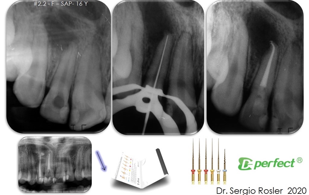
The maxillary lateral incisor apical curvature is respected after applying the shaping protocol with the T-Pro NiTi file system. (Image: Dr Sergio Rosler)
The 1-hour webinar, titled “Management of complex endodontic anatomy using PERFECT NiTi files”, will be presented live on Wednesday, 8 April, at 2 p.m. EDT. Participants will have the opportunity to ask questions about the topic as well as earn a continuing education credit by answering a questionnaire after the lecture. Registration on the Dental Tribune Study Club website is free of charge.
After access preparation and location of anatomy, the next challenge facing the endodontic clinician is to select the proper file alloy and sequence for the...
Irrigation is a major step in endodontic treatment. A variety of chemicals are used to achieve what I like to consider the chemical preparation of the ...
Root-canal anatomy with all of its inherent complexity still represents a very serious challenge to modern root-canal therapy. Even with many breakthroughs ...
From the early 20th century, when Walter Hess and Ernest Zürcher [1] demonstrated root canal anatomy with an unprecedented visual clarity, its complexity ...
Maxillary second molars are always a challenge for root canal therapy. This difficulty is related to the location of the tooth, way back in the maxilla with...
“Faster, higher, stronger”—this motto certainly no longer applies only to the Olympic Games. How can certain tasks be performed even more precisely, ...
The patient reported on in this article is a student in dentistry and his parents are both dentists. They referred their son to a good endodontist, who then...
The patient reported on in this article is a student in dentistry and his parents are both dentists. They referred their son to a good endodontist, who then...
Education
Live webinar Wed. 22 July 2026 1:00 pm EST (New York)
BREA, Calif., US: Pac-Dent, a provider of digital dentistry materials and workflow solutions, has announced the expansion of its Rodin ecosystem with the ...
MILAN, Italy: At a press briefing hosted by consumer health company Kenvue during the 2026 International Symposium on Dental Hygiene (ISDH) in Milan, ...

 Brazil / Brasil
 Canada / Canada
 Latin America / Latinoamérica
 USA / USA
 Austria / Österreich
 Bosnia and Herzegovina / Босна и Херцеговина
 Bulgaria / България
 Croatia / Hrvatska
 Czech Republic & Slovakia / Česká republika & Slovensko
 France / France
 Germany / Deutschland
 Greece / ΕΛΛΑΔΑ
 Hungary / Hungary
 Italy / Italia
 Netherlands / Nederland
 Nordic / Nordic
 Poland / Polska
 Portugal / Portugal
 Romania & Moldova / România & Moldova
 Slovenia / Slovenija
 Serbia & Montenegro / Србија и Црна Гора
 Spain / España
 Switzerland / Schweiz
 Turkey / Türkiye
 UK & Ireland / UK & Ireland
 China / 中国
 India / भारत गणराज्य
 Pakistan / Pākistān
 Vietnam / Việt Nam
 ASEAN / ASEAN
 Israel / מְדִינַת יִשְׂרָאֵל
 Algeria, Morocco & Tunisia / الجزائر والمغرب وتونس
 Middle East / Middle East


 Dr. Crystal Marruganti, Cat EdneyRegister now1CELive webinar
 Federico ZunicaRegister now1CELive webinar
 Dr. Sergio FlorencioLive webinar
 Dr. Cameron Shahbazian DMD MBARegister now1CE


To post a reply please login or register