Search Dental Tribune

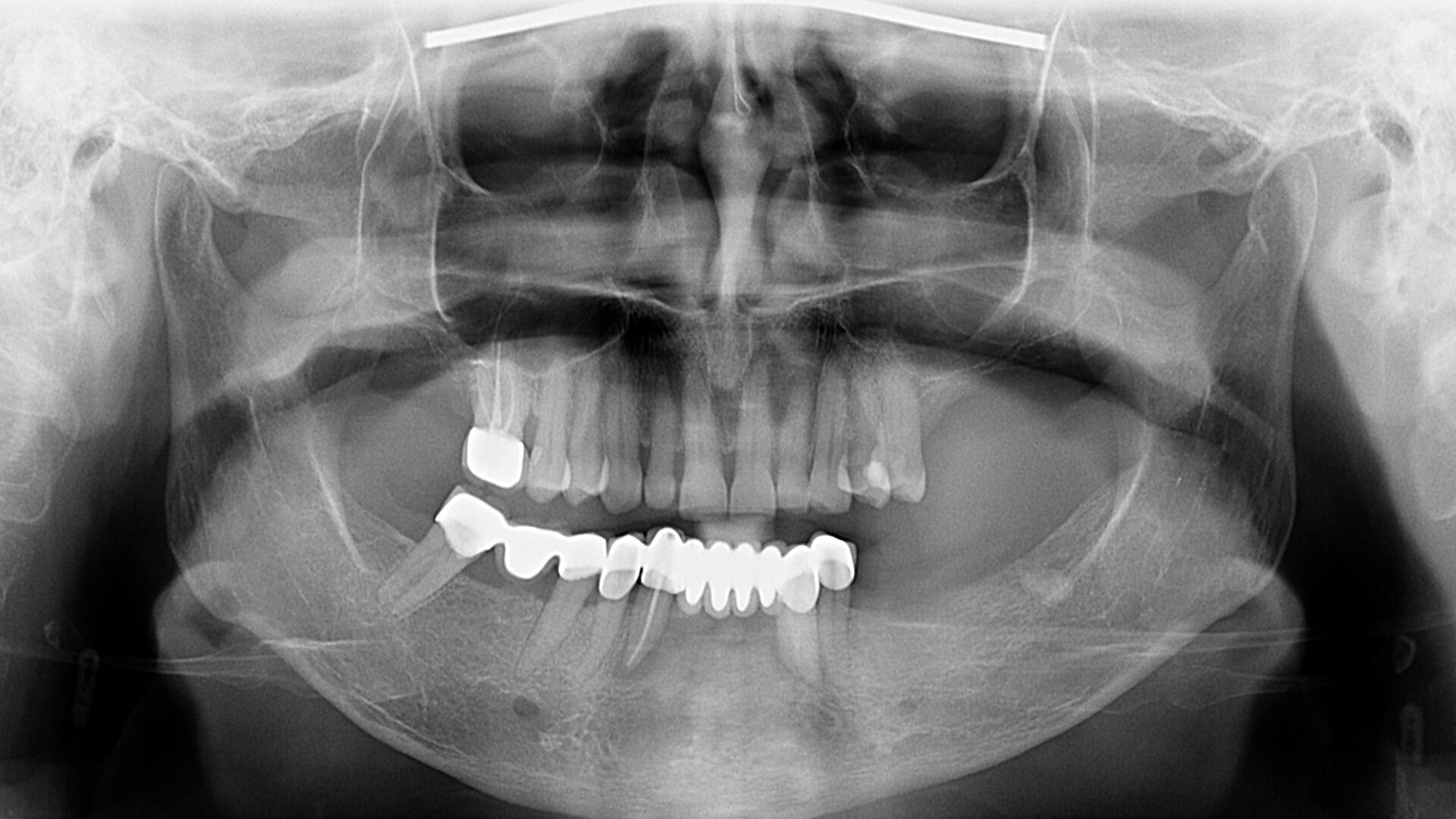
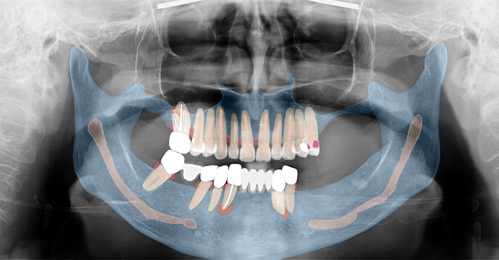
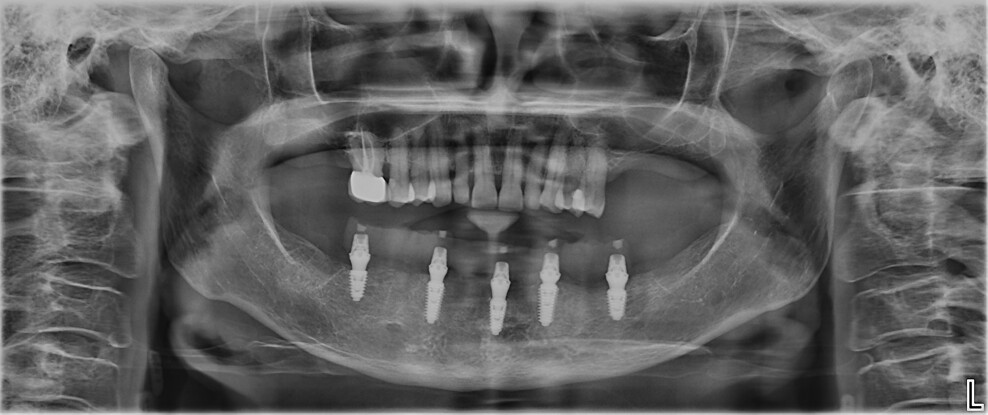
Fig. 1a: Pre-op panoramic radiograph. (All images: Dr Ádám Kapai, Dr Tamás Chikány, Tamás Csornai)

Fri. 21. November 2025

save

Modern dentistry should strive to preserve all maintainable natural teeth whenever possible, in line with the principles of minimal invasiveness and biological respect. However, in certain clinical scenarios, particularly when the remaining dentition is compromised by periodontal disease, periapical pathology and questionable long-term prognosis, full-arch rehabilitation with dental implants may represent a pragmatic, patient-driven alternative.

The all-on-X concept has evolved into a versatile treatment modality, particularly for patients who would prefer not to undergo multiple extensive hard- and soft-tissue augmentation procedures and lengthy, costly tooth-preserving therapies.1 This case report describes the diagnosis, planning and execution of a mandibular full-arch rehabilitation using an entirely digital prosthetic workflow. The case report highlights the clinical rationale behind the treatment plan options and the role of digital tools in achieving a predictable outcome.

Fig. b: Pre-op panoramic radiograph and the findings.

Fig. b: Pre-op panoramic radiograph and the findings.

Traditionally, full-arch implant rehabilitation was carried out using conventional impression techniques, requiring multiple appointments and repeated impressions and often causing discomfort for patients. Furthermore, any error during the process usually required starting over with a new impression, and this could be particularly challenging if impression copings had to be removed and repositioned. The advent of intra-oral scanners has introduced a paradigm shift in implant prosthodontics, enabling clinicians to work more efficiently and comfortably while achieving accuracy comparable to that of conventional methods.

Digital workflows have long been implemented in dental laboratories, but their integration into clinical practice in recent years has enabled a seamless transition from digital planning to definitive restoration. Initially, the accuracy of digital impressions for full-arch restorations was debated, and physical impressions were often preferred for large-span rehabilitation. However, the development of advanced scan bodies, particularly horizontal and low-profile designs, has made it possible to achieve highly accurate digital impressions even in full-arch implant cases, in both the maxilla and the mandible. These advancements, along with thoughtful material selection and planning, now allow clinicians to fully leverage the benefits of digital workflows in complex rehabilitation.

Case presentation

A 65-year-old female patient presented with complaints of tooth sensitivity under her existing mandibular prothesis and expressed concern over her missing teeth and periodontal health. The patient also inquired about the feasibility of implants to restore function and aesthetics.

Clinical and radiographic findings

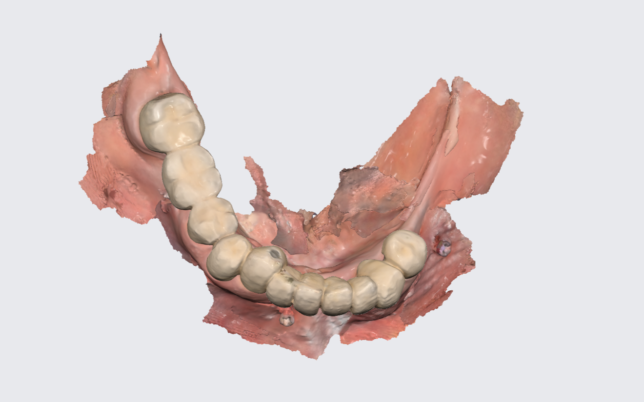
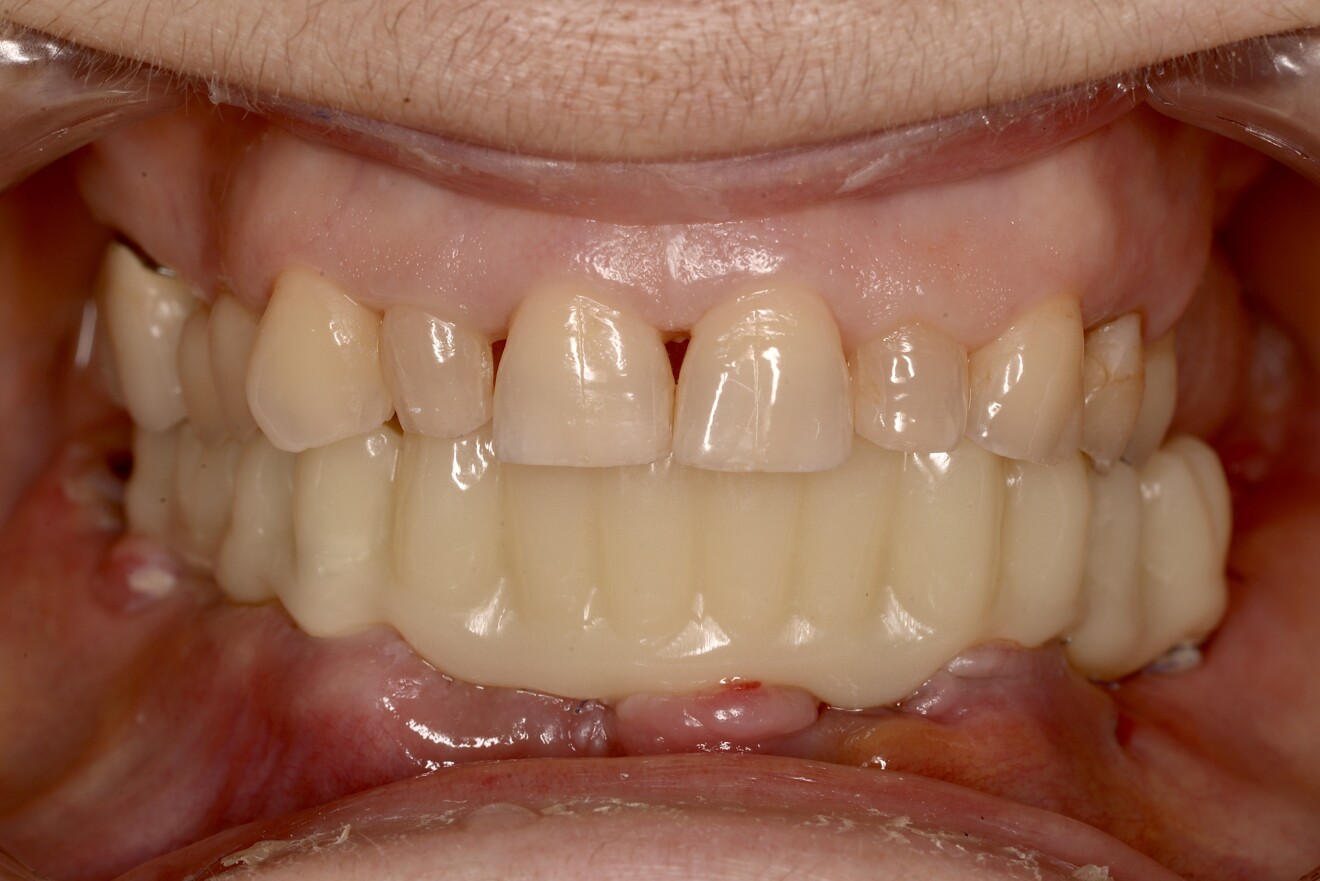
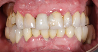
Intra-oral examination revealed compromised mandibular dentition, an old fixed prothesis, missing posterior teeth on the left side, periodontal involvement and gingival inflammation. A panoramic radiograph was obtained, and the findings were reported to the patient (Figs. 1a & b; 2a & b). Both mandibular canines had visible periapical radiolucencies. A CBCT scan was performed for a more detailed assessment of the alveolar bone anatomy (Figs. 3a & b). Temporomandibular joint examination revealed no indication of dysfunction (crepitus, clicking or pain).

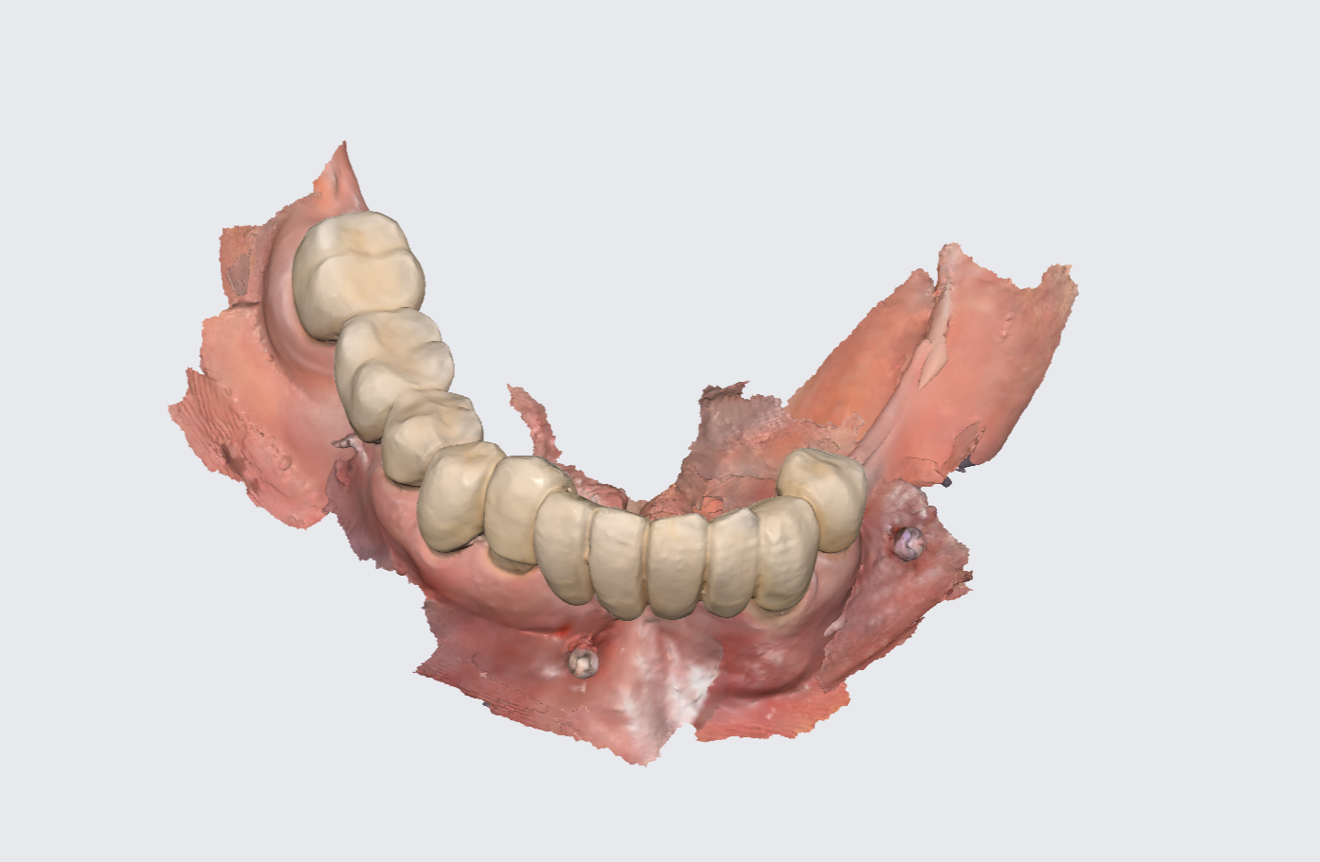
Figs. 2a & b: Intra-oral scans of the pre-op situation.

Figs. 2a & b: Intra-oral scans of the pre-op situation.

Fig. 2b

Fig. 2b

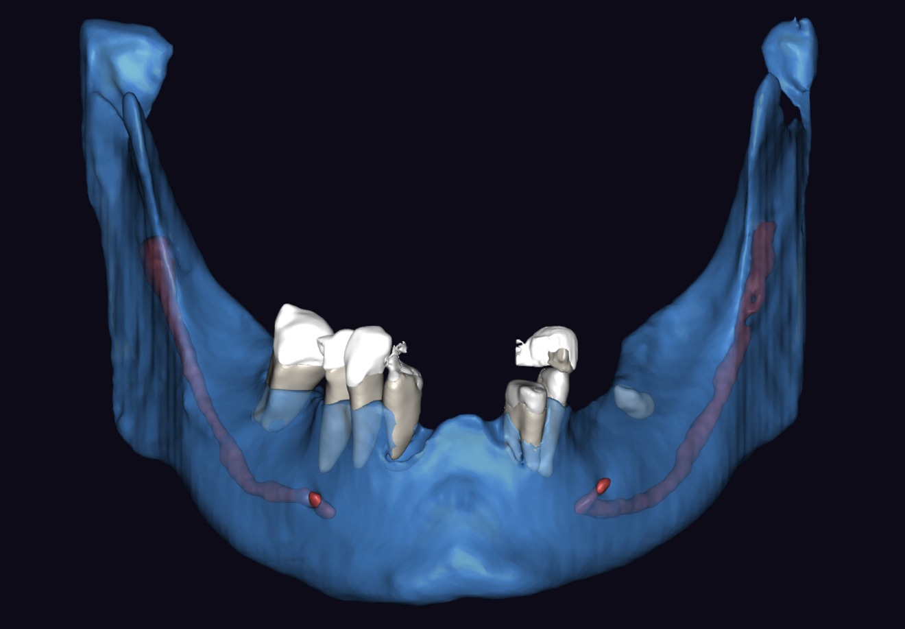
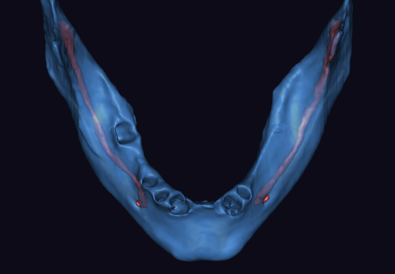
Figs. 3a & b: Segmented CBCT scan (a). Mandibular arch morphology after virtual teeth extractions (b).

Figs. 3a & b: Segmented CBCT scan (a). Mandibular arch morphology after virtual teeth extractions (b).

Fig. 3b

Fig. 3b

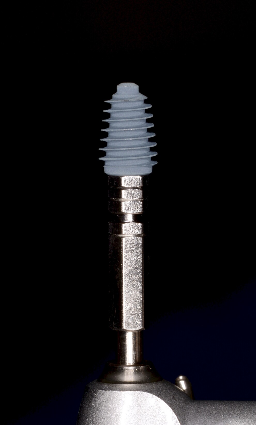
Fig. 4: AnyRidge implant (MegaGen).

Fig. 4: AnyRidge implant (MegaGen).

Treatment planning

The patient was initially offered a treatment plan aimed at preserving and utilising all salvageable teeth, complemented by strategically placed implants. This approach would have involved a comprehensive rehabilitation of the anterior and left posterior zones, requiring advanced bone grafting procedures, soft-tissue surgeries and the placement of at least four implants. This was considered the preferred treatment plan, reflecting the clinical team’s philosophy of tooth preservation whenever feasible. However, the patient declined this option owing to the longer treatment time, the need for multiple surgical interventions and the higher overall cost. As an alternative, an all-on-five full-arch rehabilitation was proposed, and this was accepted by the patient.

Surgical treatment and postoperative care

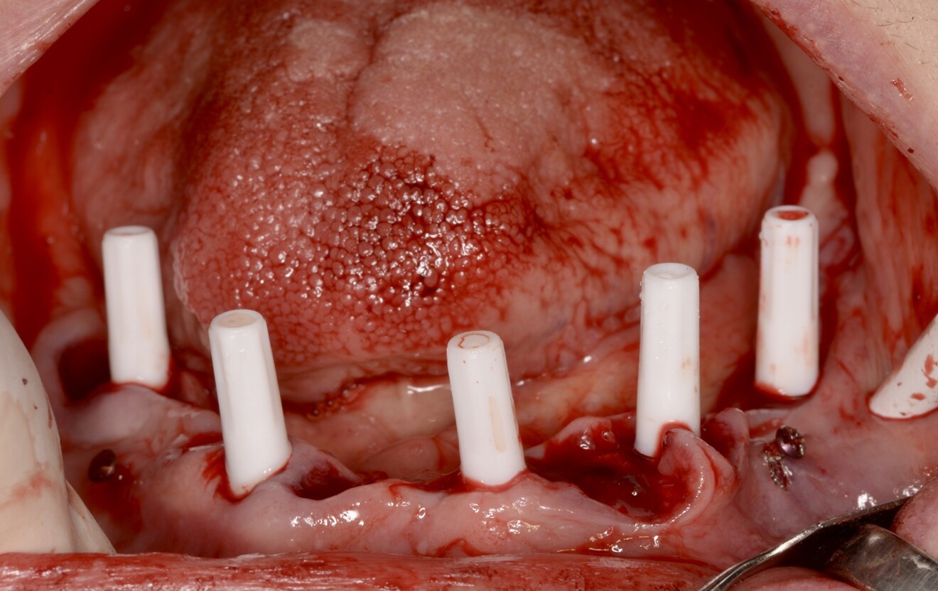
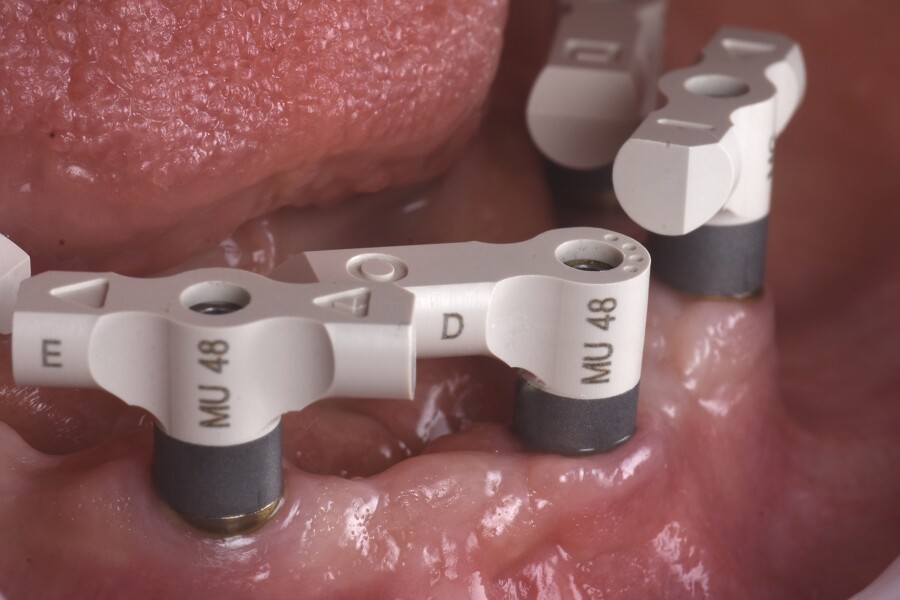
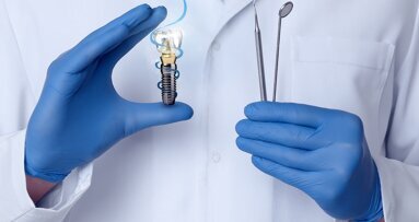
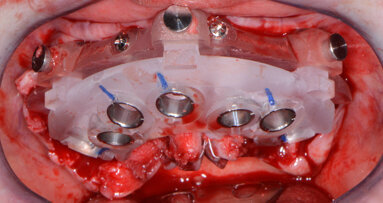
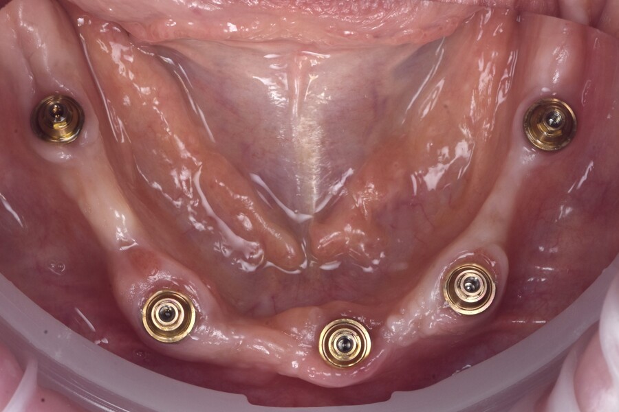
At the surgery appointment, three osteosynthesis screws were placed to facilitate digital alignment of the preoperative and postoperative scans, and the remaining mandibular teeth were extracted, followed by full-thickness flap elevation. Minimal alveolar bone reduction was performed to improve ease of cleaning and to position the transitional line more apically. Five AnyRidge implants (MegaGen) were placed sub-crestally in native bone, avoiding the need for bone grafting (Fig. 4). Straight N-type multi-unit abutments (MegaGen) were connected and torqued to 35 N cm. The further prosthetic workflow was carried out entirely at the abutment level, avoiding implant–abutment disconnections and thereby simplifying the prosthetic procedures (Fig. 5). This approach is favourable for crestal bone stability.

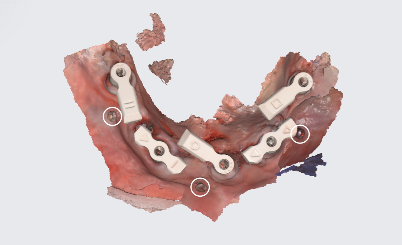
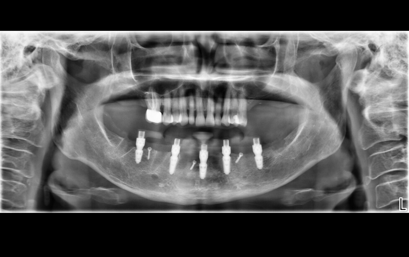
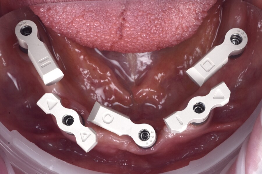
Sufficient primary stability was achieved, enabling immediate loading, consistent with evidence-based recommendations.2–6 SmartFlag scan bodies (Apollo Implant Components) were attached, and the postoperative intra-oral scan was performed with both the scan bodies and the osteosynthesis screws in place (Figs. 6 & 7). The screws were removed after scanning.

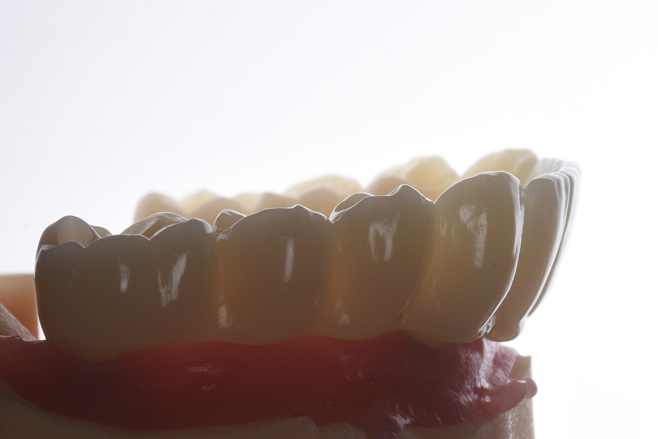
Postoperative medication consisted of a seven-day course of amoxicillin–clavulanic acid (875/125 mg, twice daily), diclofenac (50 mg, up to three times daily as needed) and a chlorhexidine mouthrinse (two to three times daily). The patient was recalled the following day for delivery of the screw-retained PMMA provisional restoration, which showed excellent passive fit and acceptable occlusion (Figs. 8a & b).

At the one-week follow-up, the sutures were removed, the prosthesis was re-evaluated clinically and radiographically, and the occlusion was reassessed. The patient adapted easily to the restoration. Oral hygiene instructions were given, and the patient’s excellent baseline oral hygiene and motivation were noted as positive prognostic factors.

Fig. 5: Intra-op photograph after attachment of the osteosynthesis screws and implant placement.

Fig. 5: Intra-op photograph after attachment of the osteosynthesis screws and implant placement.

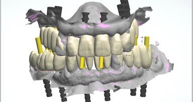
Fig. 6: Post-op scan with SmartFlags and osteosynthesis screws (circled).

Fig. 6: Post-op scan with SmartFlags and osteosynthesis screws (circled).

Fig. 7: Panoramic radiograph for scan body verification.

Fig. 7: Panoramic radiograph for scan body verification.

Figs. 8a: Screw-retained PMMA provisional restoration.

Figs. 8a: Screw-retained PMMA provisional restoration.

Fig. 8b: Delivery of the provisional restoration the day after the surgery.

Fig. 8b: Delivery of the provisional restoration the day after the surgery.

Digital workflow for screw-retained provisional restoration

Fully digital full-arch rehabilitation presents unique challenges, particularly in the mandible, where the absence of stable anatomical landmarks and soft-tissue changes after extractions and alveoloplasty can compromise accurate alignment of preoperative and postoperative scans. This digital workflow was designed with these challenges in mind to ensure a predictable and efficient process.

Fig. 9: Panoramic radiograph after five months of healing.

Fig. 9: Panoramic radiograph after five months of healing.

Preoperatively, three osteosynthesis screws were placed as fiducial markers. These screws provided fixed, easily identifiable reference points on the radiographs and digital scans, enabling precise alignment of the intra-oral scans before and after surgery. This is critical in the mandible, where no equivalent of the palatal rugae is available and where soft-tissue contours change significantly after flap elevation and suturing.

The use of SmartFlag scan bodies intra-operatively further enhanced workflow accuracy. Compared with conventional vertical scan bodies, the SmartFlag’s compact, horizontal geometry and clearly defined reference surfaces enable superior scanner recognition and reduce scanner signal loss. The low-profile shape of SmartFlag minimises the need for frequent repositioning of the scanner and limits cumulative errors often associated with scanning long-span restorations. These properties were particularly advantageous in this full-arch mandibular case, enabling accurate digital capture of the implant positions. Postoperatively, a single intra-oral scan with the SmartFlag scan bodies was performed and digitally aligned with the preoperative scan using the fiducial markers. This facilitated the design and delivery of the immediately loaded provisional restoration with a passive, well-adapted fit. This protocol underscores the importance of preoperative planning and the integration of advanced tools like SmartFlag scan bodies to achieve predictable, reproducible outcomes in fully digital full-arch rehabilitation.

Final prosthetic workflow

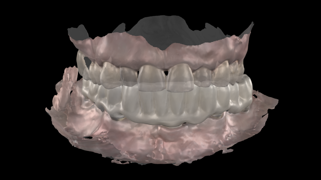
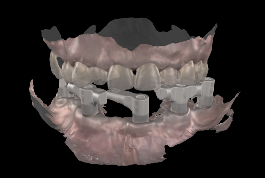
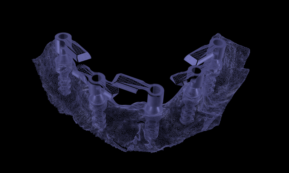
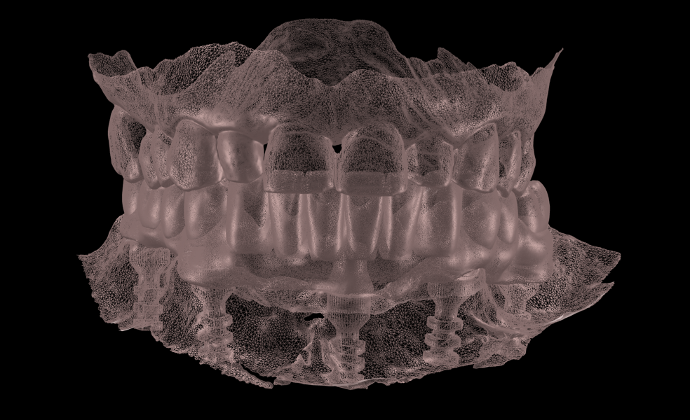
After a five-month healing period, fabrication of the definitive restoration was initiated (Figs. 9 & 10). The Medit SmartX workflow was employed, though the scan body matching was performed manually rather than using the software’s automatic feature (Figs. 11a & b). Three intra-oral scans were acquired: a scan of the SmartFlag scan bodies, a scan of the edentulous mandibular ridge and a scan of the provisional restoration in place. These datasets were aligned to replicate the occlusal scheme and soft-tissue contours (Figs. 12a & b; 13a & b).

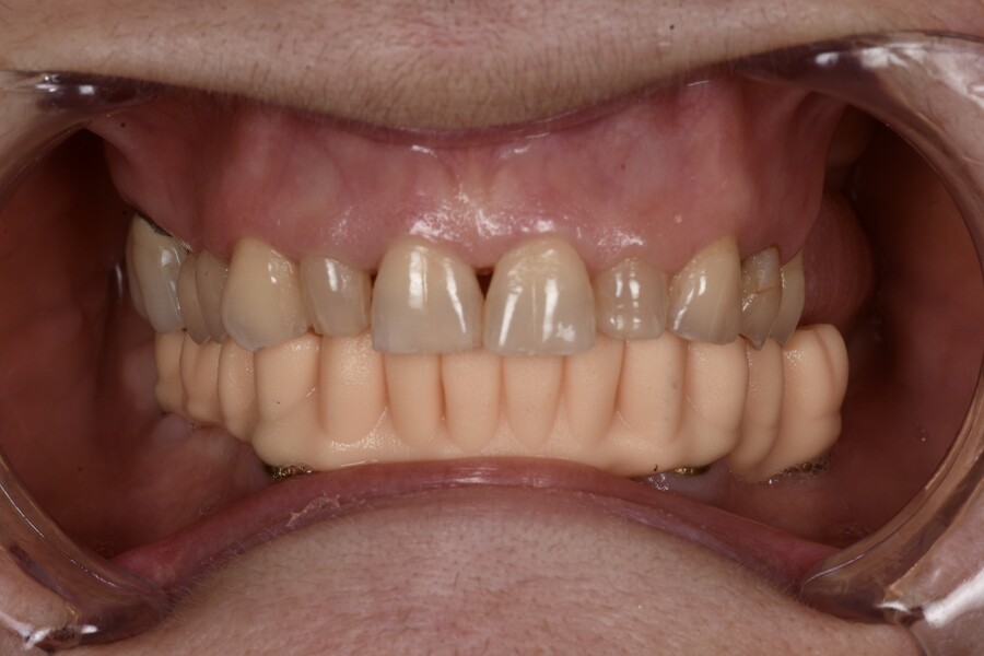
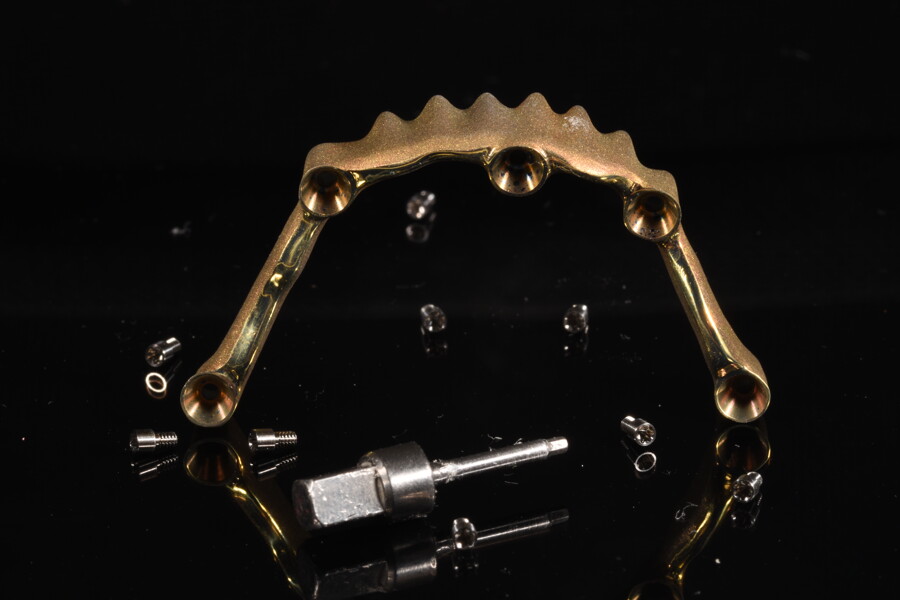
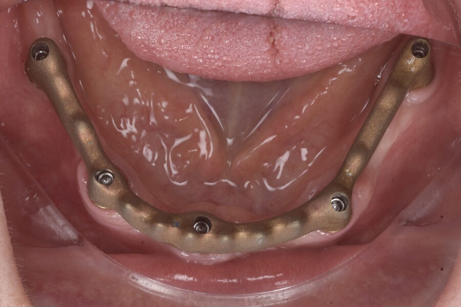
A 3D-printed diagnostic prosthesis was tried in to assess fit and occlusion; and minor occlusal adjustments were made (Fig. 14). The definitive prosthesis was fabricated as a Misch classification FP-3 screw-retained restoration, replacing missing teeth and compensating for alveolar soft-tissue deficits with an aesthetically designed gingival component.7 A titanium bar was then digitally designed and milled to directly engage the multi-unit abutments without the need for titanium bases.

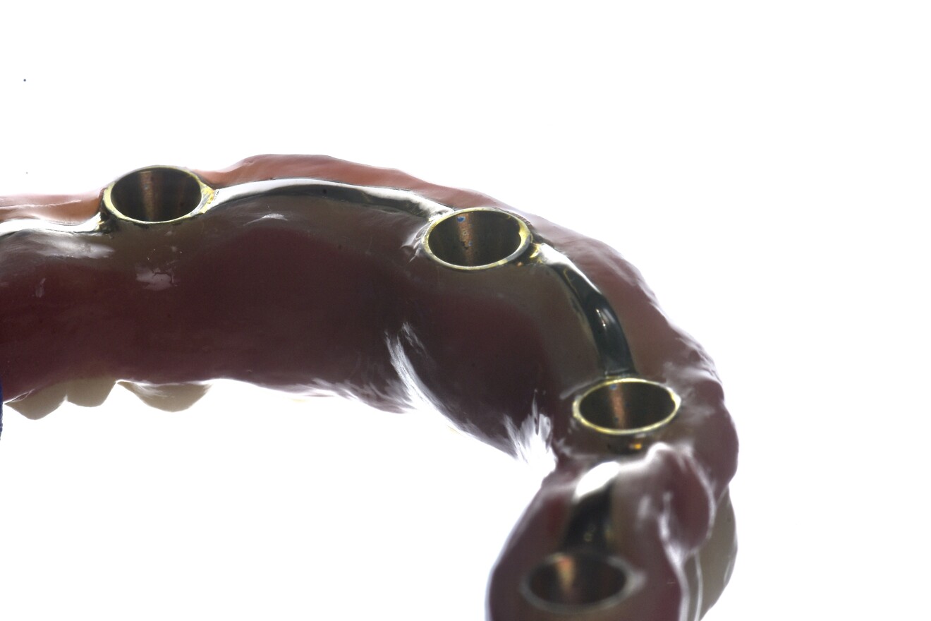
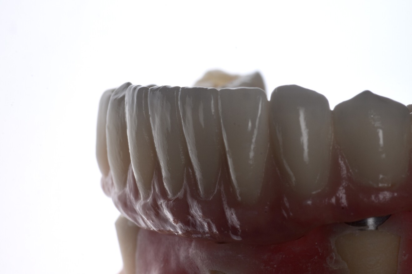
The titanium bar was anodised for improved aesthetics, producing a warm, gold-like hue that harmonised better with the gingival ceramic and teeth. Anodisation may also enhance surface roughness and increase bond strength to polymer-based or ceramic superstructures, although the evidence in the context of full-arch restorations remains limited.8 The titanium bar was tried in separately, and a modified Sheffield test confirmed passive fit (Figs. 15a & b). A monolithic full-contour zirconia superstructure was bonded on to the anodised titanium bar in the laboratory using a resin-based luting cement.9 The gingival portion of the zirconia was customised with pink ceramic. All the screw access holes were straight; however, the Apollo Implant Components’ prosthetic system is capable of up to 36° of angulation, providing prosthetic flexibility and allowing for optimised location of access holes, a capability that can significantly improve aesthetics, particularly in the maxillary anterior zone.

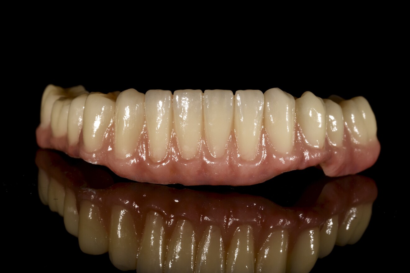
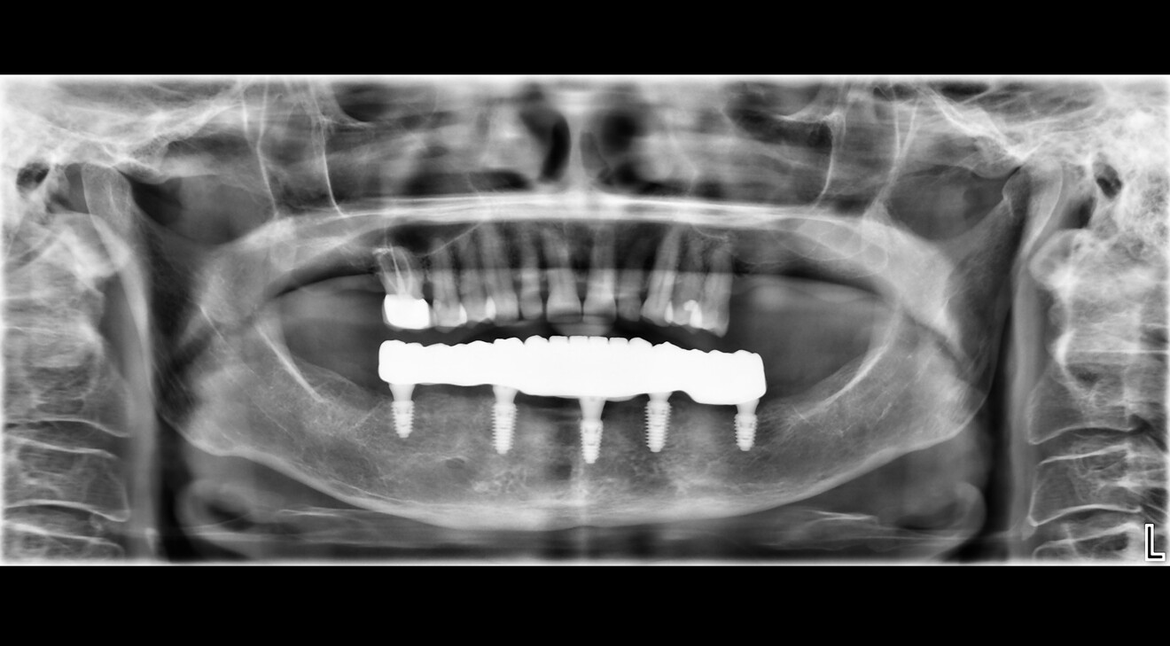
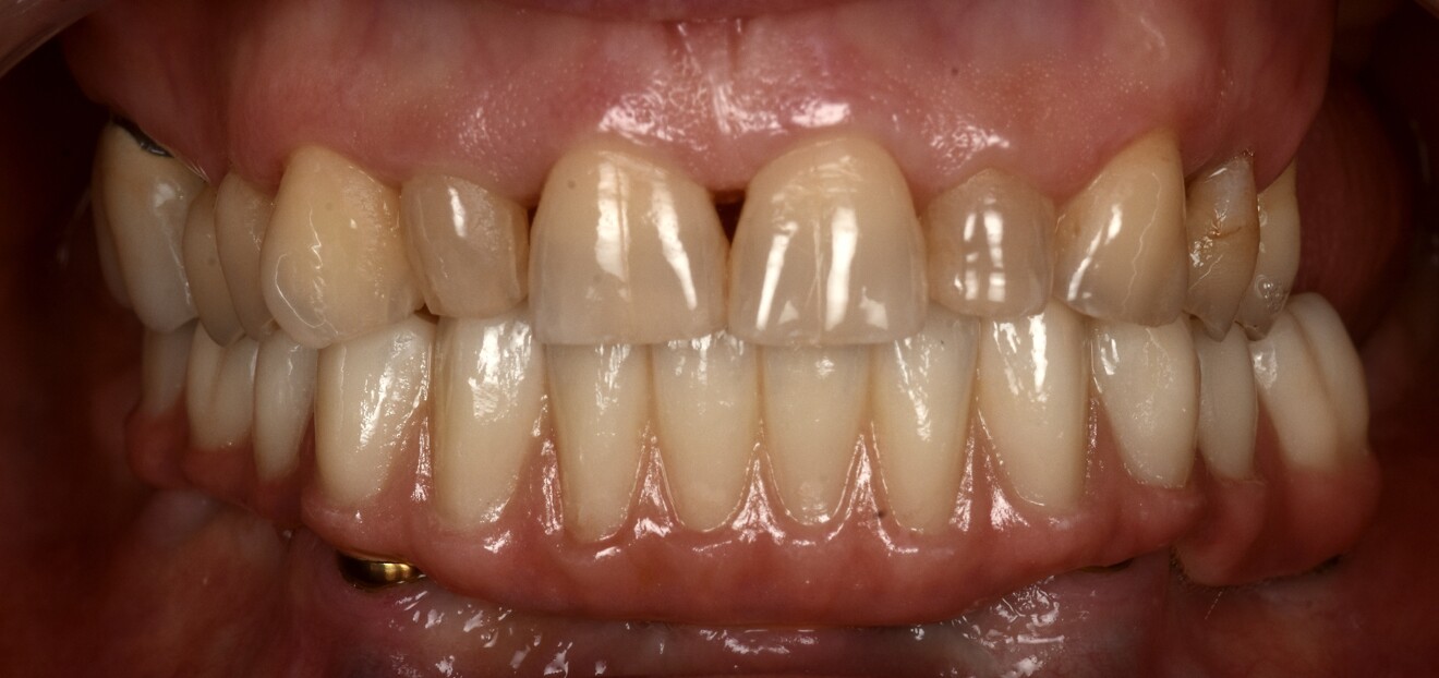
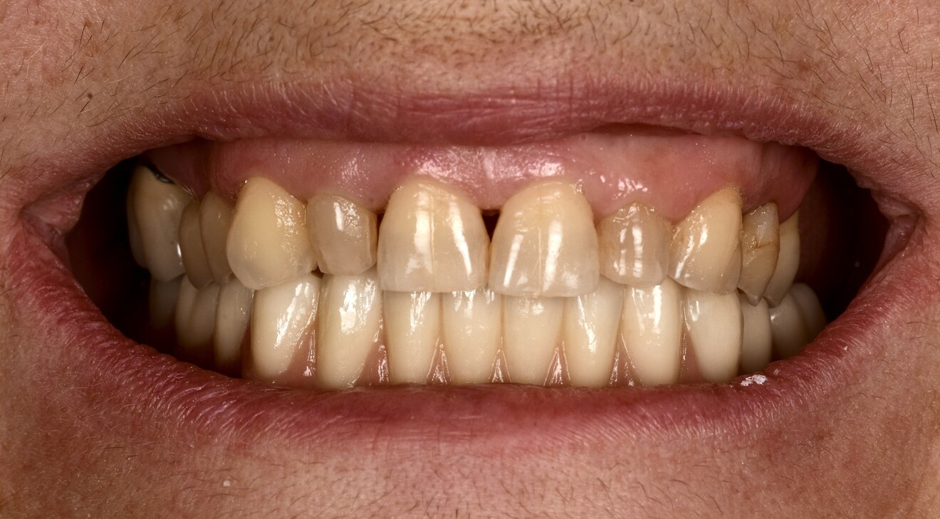
At the final delivery appointment, the definitive prosthesis was placed. A final radiograph was obtained to verify seating and fit, and then the prosthesis was torqued to 15 N cm as recommended. The occlusion and articulation were checked and adjusted as necessary. The screw access holes were sealed with PTFE tape and Shade A3 flowable composite (Figs. 16a-f). The patient was then instructed on hygiene and maintenance.

Figs. 16a–c: Definitive monolithic zirconia prothesis bonded to the anodised titanium bar (a–c).

Figs. 16a–c: Definitive monolithic zirconia prothesis bonded to the anodised titanium bar (a–c).

Fig. 16b

Fig. 16b

Fig. 16c

Fig. 16c

Fig. 16d: Final panoramic radiograph.

Fig. 16d: Final panoramic radiograph.

Fig. 16e: Final intra-oral view.

Fig. 16e: Final intra-oral view.

Fig. 16f: Final frontal view of the patient’s smile.

Fig. 16f: Final frontal view of the patient’s smile.

Discussion

A notable consideration in this case was the clinically healthy and asymptomatic condition of the temporomandibular joints. While centric relation is commonly preferred in full-mouth rehabilitation owing to its reproducibility and stability, maintaining maximal intercuspal position was chosen in this case. This decision was based on the absence of maxillary restoration and the patient’s asymptomatic temporomandibular joints. Avoiding unnecessary alteration to the patient’s habitual occlusion provided easier adaptation and comfort, aligning clinical prosthodontic principles with patient-specific conditions and preferences.10, 11

Another aspect worth highlighting is the decision to perform the prosthetic workflow entirely at the abutment level. This approach aligns with the one abutment, one time concept, avoiding repeated implant–abutment disconnections and minimising the disruption of hemidesmosomal attachment. The multi-unit platform facilitates prosthetic passivity, because implant angulations are less critical when using multi-unit abutments. Higher angulation differences can be compensated for with angled multi-unit abutment as well. The multi-unit platform is designed for ease of use, simplifying scanning, impression taking, try-in and final delivery. It also allows the prosthetic platform to sit more coronally, facilitating easier access for evaluation of fit and passivity. For this particular implant system, implant-level restorations are contra-indicated owing to the Morse taper connection, which makes it challenging to seat multi-implant prostheses passively when axes diverge. Thus, an abutment-level workflow provides mechanical, biological and practical advantages in full-arch rehabilitation.

The use of fiducial markers in this case also deserves mention. While alternatives such as adhesive markers or digitally designed tissue trackers have been described, the principle remains the same: stable, scanner-visible reference structures should be placed before surgery that remain unchanged throughout the procedure. This ensures easily identifiable landmarks for aligning preoperative and postoperative data, improving the accuracy of digital workflows in full-arch cases.

The material selection in this case was also a key consideration. A titanium bar supporting a monolithic zirconia superstructure was chosen over a full-zirconia or metal–ceramic restoration. This approach combined strength, aesthetics and long-term reliability. While full-zirconia restorations are becoming increasingly popular for their aesthetic qualities and structural durability as monolithic restorations, they are prone to fracture if passive fit is compromised, particularly in the mandible owing to its inherent flexure. Conversely, metal–ceramic restorations, while robust, lack the same level of translucency and natural aesthetics and are not suitable for a fully digital workflow, although the framework could be used in the CAD/CAM digital workflow. The titanium bar provided a strong, stable framework, while the zirconia superstructure offered superior aesthetics and biocompatibility. This combination represents a balanced solution tailored to the clinical demands and patient expectations of full-arch rehabilitation.

This case demonstrates the importance of a carefully planned, fully digital workflow in full-arch mandibular rehabilitation. Beyond accuracy and predictable fit, the fully digital approach offers increased efficiency, fewer clinical sessions and improved patient comfort compared with conventional impressions. The ability to replicate the provisional restoration’s occlusion and use its design—so-called copy–paste dentistry—further simplifies and streamlines the process.12 Digital workflows also contribute to improved communication between the clinical and laboratory teams and allow for earlier patient function and satisfaction. Importantly, the workflow followed in this case shows that modern digital techniques can deliver outcomes comparable to those of traditional methods and offer added benefits in workflow speed and convenience. Fully digital workflows of this kind highlight the clinical value of the approach.

Conclusion

This case demonstrates how a fully digital workflow can facilitate a predictable, patient-centred full-arch rehabilitation in the mandible, even when tooth-preserving treatment is declined. Careful planning, the use of fiducial markers and SmartFlag scan bodies, and adherence to prosthetic principles allowed the team to achieve a passive, functional and aesthetic restoration. Digital workflows offer promising alternatives to conventional techniques, provided their limitations are understood and mitigated.

Editorial note:

This article was published in digital—international magazine of digital dentisty vol. 6, issue 3/2025. The list of references can be found here.
Topics:
Tags:
To post a reply please login or register
advertisement
advertisement