The purpose of this article is to show the diferent modalities of treatment for upper lateral incisors agenesis. In daily practice, orthodontists often meet cases of upper lateral incisors agenesis. The two common treatment options are: space closure, using canines to substitute the missing lateral incisors, or space opening for future restorations.
Introduction
- Theoretical considerations.
- The second most common agenesis, representing 20 % of the congenital missing teeth.
- The second most common agenesis (Europe), after the mandibular second premolar.
- The most frequently missing tooth in the American population.
- Unilateral agenesis is often associated with dysmorphia or microdontia of the corresponding contralateral tooth.
Treatment alternatives
- Space opening for future restorations.
- Space closure–canine substitution of the lateral incisor.
Treatment objectives
- Optimal dentogingival aesthetics.
- Functional occlusion.
Optimal dentogingival aesthetics objectives
- Gingival height of contour of the upper anterior teeth (Fig. 1): central incisors and canines are more superior than that of the lateral incisors.
- The long axis of the central incisor and the canines should be slightly mesial to the gingival height of contour (Fig. 2).
- The long axis of the upper lateral incisor should be coincidental to the gingival height of contour (Fig. 2).
- Dental proportions: the width of wellproportioned teeth should be approximately 60 % to 75 % of their height (Fig. 3).
Functional occlusion objectives
- 3–4 mm of overbite.
- 0–2 mm of overjet.
- Anterior and canine guidance, which allows for the immediate disclusion of molars and premolars when making lateral or protrusive movements.
- Centric occlusion coinciding with centric relation.
Treatment strategies for canine substitution
- Angulate and extrude the canine to mimic an upper lateral incisor relative to the gingival height of contour.
- Intrude the upper first premolar to mimic an upper canine relative to the gingival height of contour.
- Apply lingual root torque to mimic the emergence profile of the lateral incisor and improve the emergence profile of the bulky gingival tissue of the substituted canine: use a lower second premolar bracket on the upper canine.
- Adjust dental proportions as necessary: mesiodistal reductions on the upper central incisors to balance adjustments on the substituted canine.
When is canine substitution appropriate?
Occlusal considerations
- Class II free of mandibular crowding: molars in full Class II and premolar brought forward to act as the canine, while remaining in a Class I relationship with the lower canine.
- Class I with sufficient mandibular anterior crowding that would necessitate premolar extractions on the lower arch.
Profile
- Flat.
- Slightly convex profile.
Canine size, shape and colour
- The width at the cementoenamel junction: the wider the tooth, the more difficult it will be to mimic a lateral incisor.
- Colour: canines are the teeth that are the most saturated with chroma. A canine that is smaller in shape and does not have an oversaturation of chroma would make an excellent candidate for canine substitution.
Smiling lip level
- Depending on how high the smile line is, it may show the canine eminence.
- Large canines often have an obvious root prominence, and high lip levels may reveal that there is an unnatural eminence in the lateral sight.
Clinical case
A 13-year-old patient complaining about the aesthetic aspect of her smile was sent to my office by a general dentist with a diagnosis of the bilateral lateral incisors agenesis.
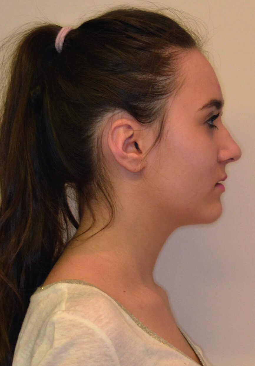
The treatment started with an aesthetic analysis of the patient’s face (Figs. 4–10), which was as follows:
- Square face.
- Slight facial asymmetry, with menton deviated to the right.
- Maxillomandibular biretrusion (Fig. 10).
- Correct curl of the upper lip.
- Left side of the face is more flat compared with the right side.
- Right eye slightly higher.
- Insuficient display of the upper anterior teeth with lips in repose.
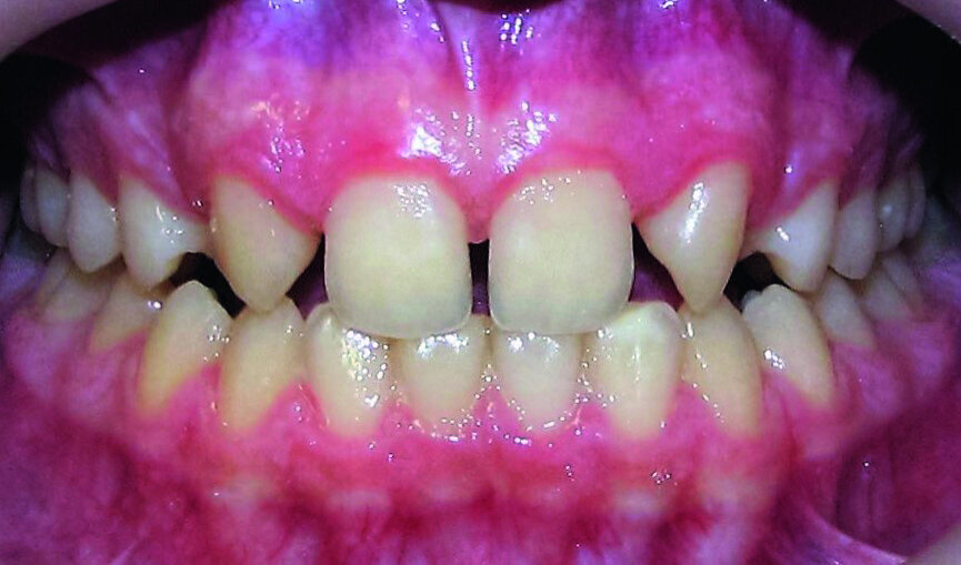
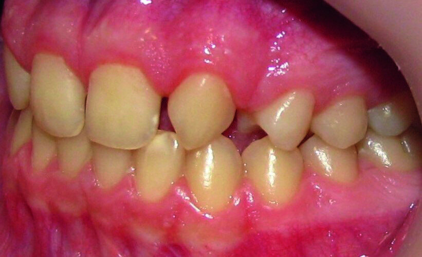
Then occlusal analysis (Figs. 11–21) was performed:
- Skeletal Class III (Fig. 17).
- Dental Class I.
- Maxillary hypoplasia with lower arch dental compensations.
- Insuficient overbite and overjet.
- Upper spacing due to the laterals agenesis (Fig. 18).
- Upper midline deviated to the right.
- Initial CR mounting (Figs. 19–21).
Finally space analysis was carried out (Tables 1a & b).
Treatment plan
It was concluded that maxillary hypoplasia was an indication for space opening. Considering the young age of the patient, the skeletal pattern and the high demands regarding aesthetics, it was decided to:
- Open spaces for two implants, but in the posterior area: 14 and 24.
- Substitute the laterals with canines.
- Temporary implants and crowns on teeth #14 and 24, until 18 years.
- Reshaping the canines and first premolars to match the shape of the lateral incisors and canines: direct composites restorations on teeth #13, 11, 21, and 23.
Treatment step by step:
- Upper bracket placement (Fig. 22): level and align the gingival margins, and correct the torque on the upper canines who will substitute the lateral incisors.
- Implants space opening: substitute laterals with canines (Figs. 23–25) and substitute canines with first premolars (Figs. 26–28).
- Finishing and occlusal settling (Figs. 29–31).
- Verifying the implant site width (Fig. 32) and provisory implants and crowns placement (Fig. 33).
Restorative phase
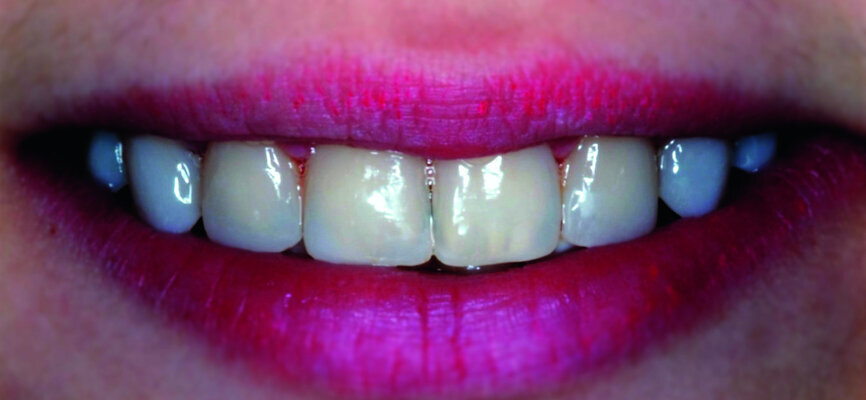
At the end of orthodontic treatment, gingival margins were well aligned, midlines were centred and canines and first premolars were positioned to facilitate the restorative phase of treatment (Figs. 34–36). At this phase, a final CR mounting and wax-up was performed (Figs. 37–39), as well as the anterior teeth restorations (Fig. 40).
Orthodontic treatment has improved both dental and facial aesthetics (Figs. 41–47) and the functionality of the occlusion (Figs. 48–52).
Conclusions
Canine substitution can be an excellent treatment alternative for congenitally missing maxillary lateral incisors. Patient selection is critical and depends on the type of malocclusion, profile, canine shape and colour, and smile lip level. Pre-treatment evaluation of these selection criteria is necessary to ensure treatment success and predictable aesthetics.
When planning to replace congenitally missing lateral incisors, you should remember that an interdisciplinary approach is necessary to provide the most predictable treatment outcome.
The orthodontist should always consider the patient’s age, skeletal and facial pattern, dentoalveolar crowding, as well as performing dental and facial aesthetic analyses.
Editorial note: A list of references is available from the publisher. This article was published in ortho - international magazine of othodontics No. 02/2017.
The second most common dental agenesis is that of the maxillary lateral incisors, after agenesis of the mandibular third molars.1 This common agenesis has ...
Faced by a missing lateral incisor, practitioners often consider a wide range of issues and are also faced by numerous treatment options:
1. in a young ...
Having discussed in the previous article (see editorial note), all of the preprosthetic stages for the replacement of a lateral maxillary incisor and having...
The absence of the maxillary lateral incisors creates a functional and esthetic problem that can be managed with different treatment modalities.
Minimally invasive treatments restore form, function and aesthetics with minimal removal of sound tooth structure. Understandably, the restorations age with...
Placing an implant immediately after tooth extraction offers several advantages, including a reduced treatment time, fewer surgical sessions, possibility of...
Numerous clinical studies confirm that maxillary transverse deficiency is among the most common orthodontic and skeletal problems in childhood and is ...
Porcelain-fused to metal crowns have been commonly used since their introduction in the 1960s.1 The presence of a metallic substructure confers certain ...
Maxillary lateral incisor agenesis (MLIA) is a congenital condition in which at least one of the maxillary lateral incisors is missing in the primary or ...
Maxillary second molars are always a challenge for root canal therapy. This difficulty is related to the location of the tooth, way back in the maxilla with...
Live webinar
Wed. 22 July 2026
1:00 pm EST (New York)
Live webinar
Thu. 23 July 2026
2:00 pm EST (New York)
Dr. Crystal Marruganti, Cat Edney
Live webinar
Thu. 23 July 2026
2:00 pm EST (New York)
Live webinar
Mon. 27 July 2026
4:00 am EST (New York)
Live webinar
Fri. 31 July 2026
12:00 pm EST (New York)
Live webinar
Mon. 3 August 2026
8:00 pm EST (New York)
Dr. Cameron Shahbazian DMD MBA
Live webinar
Wed. 5 August 2026
1:00 pm EST (New York)

 Brazil / Brasil
 Canada / Canada
 Latin America / Latinoamérica
 USA / USA
 Austria / Österreich
 Bosnia and Herzegovina / Босна и Херцеговина
 Bulgaria / България
 Croatia / Hrvatska
 Czech Republic & Slovakia / Česká republika & Slovensko
 France / France
 Germany / Deutschland
 Greece / ΕΛΛΑΔΑ
 Hungary / Hungary
 Italy / Italia
 Netherlands / Nederland
 Nordic / Nordic
 Poland / Polska
 Portugal / Portugal
 Romania & Moldova / România & Moldova
 Slovenia / Slovenija
 Serbia & Montenegro / Србија и Црна Гора
 Spain / España
 Switzerland / Schweiz
 Turkey / Türkiye
 UK & Ireland / UK & Ireland
 China / 中国
 India / भारत गणराज्य
 Pakistan / Pākistān
 Vietnam / Việt Nam
 ASEAN / ASEAN
 Israel / מְדִינַת יִשְׂרָאֵל
 Algeria, Morocco & Tunisia / الجزائر والمغرب وتونس
 Middle East / Middle East


 Dr. Crystal Marruganti, Cat EdneyRegister now1CELive webinar
 Federico ZunicaRegister now1CELive webinar
 Dr. Sergio FlorencioLive webinar
 Dr. Cameron Shahbazian DMD MBARegister now1CE


: An interdisciplinary approach to missing maxillary lateral incisors")


To post a reply please login or register