Search Dental Tribune

Surgical extrusion with 180° rotation: A case report with four-year follow-up

Surgical extrusion provides clinicians with a viable option to preserve compromised teeth and achieve stable, aesthetic long-term outcomes. (Image: Georgii/Adobe Stock; clinical images: Dr Jenner Argueta)

In recent years, dental implants have emerged as a pivotal advancement in dentistry, offering a popular solution for replacing natural teeth lost owing to various factors, such as caries, fractures and periodontal disease. However, within the dental community, there remains a divergence in approaches, some advocating for a more conservative stance focused on preserving severely compromised teeth.1 Prioritising the retention of a patient’s natural teeth should remain a fundamental objective for dental professionals.

One proposed method to address structurally compromised teeth is crown lengthening, aimed at providing optimal conditions for tooth restoration. However, challenges arise with this approach, particularly in the aesthetic zone, where certain techniques may be limited by their impact on gingival symmetry.2, 3 The diagnostic and planning process for crown lengthening of anterior teeth is notably intricate, necessitating careful consideration to avoid potential asymmetry in gingival margins. Particularly prevalent in the aesthetic zone, notably the maxillary incisors, dentoalveolar trauma often results in the loss of coronal structure.4, 5 In such cases, restoration of the affected tooth may require additional procedures to achieve adequate supragingival height.

Surgical extrusion is a procedure through which the remaining dental structure is repositioned in a more coronal position within the alveolus.6–8 The primary objective of this technique is to elevate the affected tooth to a more coronal position, thereby creating favourable conditions for establishing an adequate ferrule, crucial for facilitating the placement of a restoration that preserves a healthy biological width.3, 7 Consequently, surgical extrusion can be a valuable approach in the treatment of severely damaged teeth, particularly in the aesthetic zone.

Surgical extrusion has various names, including intra-alveolar transplant, intentional reimplantation and forced eruption.6, 9, 10 Although the technique was initially described in 1978,11 the first case report was not published until 2002.12 Despite its early documentation, surgical extrusion remains relatively uncommon in dental practice.

Initially, the periodontal ligament is delicately loosened through syndesmotomy, followed by careful luxation facilitated by periotomes and/or elevators. Using forceps, the tooth is then gradually extruded, typically achieving a vertical displacement of 4–6 mm. To stabilise the tooth in its new position, it is immobilised for a period of two to three weeks with the aid of a flexible splint, followed by placement of a post and core and subsequently a definitive full-coverage restoration. This effective technique can be implemented with relative ease, requiring no specialised surgical expertise. Moreover, it often yields satisfactory aesthetic results, boasts a low failure rate and tends to be well received by patients.

Case series and clinical reports are classified as low-evidence literature because the causal relationships between intervention and outcomes cannot be definitively established without a control group.13, 14 Nonetheless, clinical reports can influence decision-making in dental practice,14 for example by raising awareness of new techniques, clinical approaches and research directions. The objective of this case report is to present a four-year follow-up clinical case in which a compromised anterior tooth was preserved through surgical extrusion.

Case report

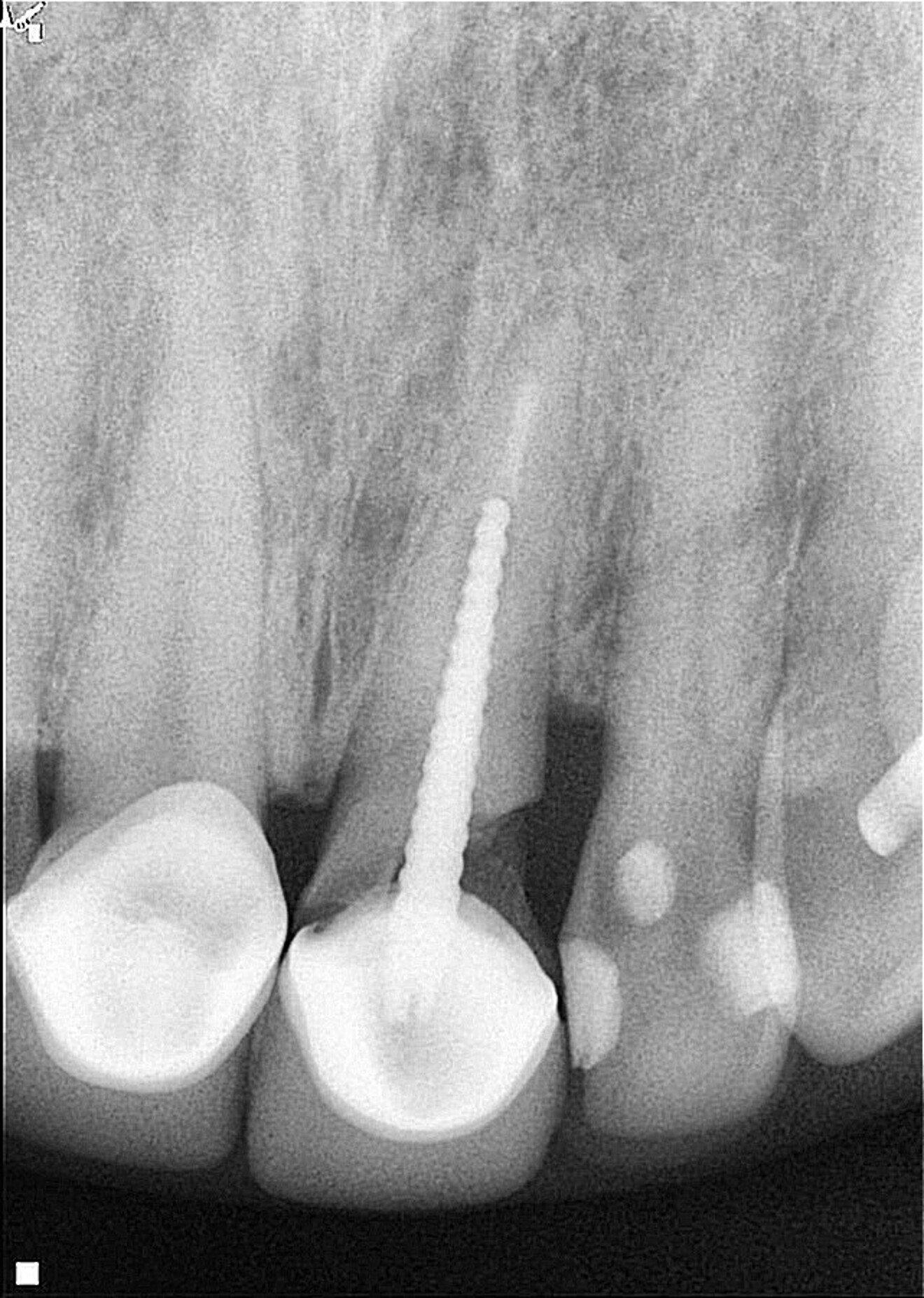
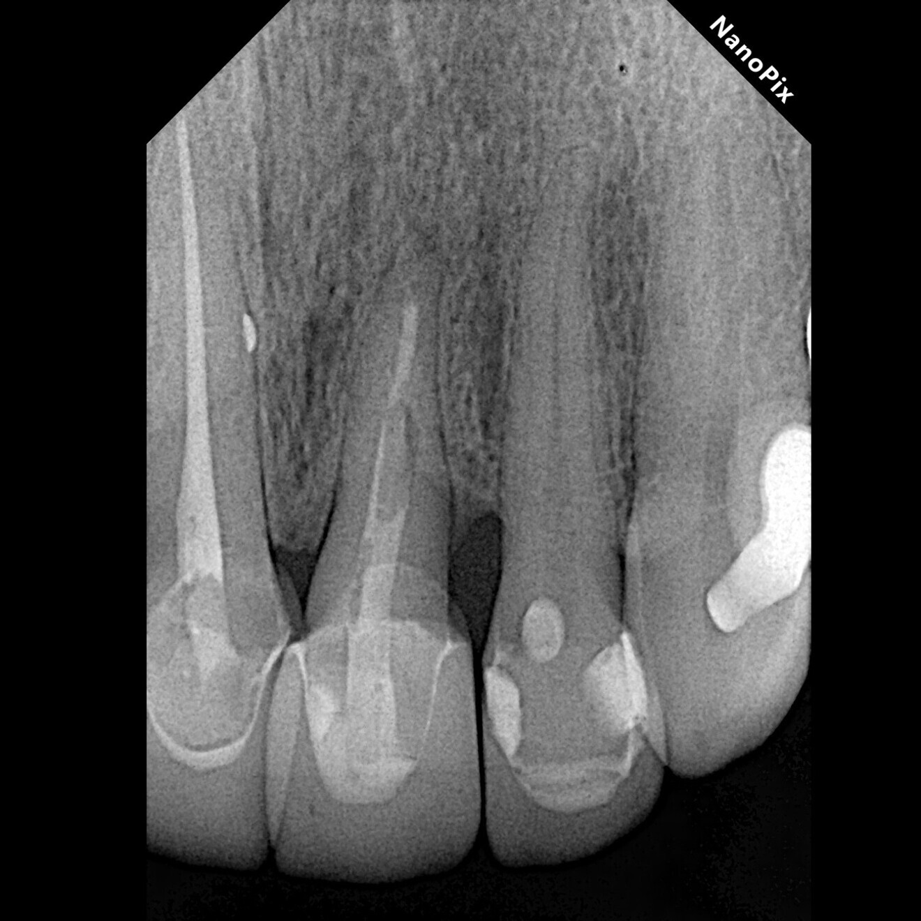
A female patient was evaluated in the dental office after a traumatic event involving her maxillary teeth. She had an oblique fracture from the mesial to distal aspect of the maxillary left central incisor at subgingival level, and the tooth’s metal post and metal–porcelain crown presented with mobility. The tooth had healthy periapical tissue (Fig. 1). Upon radiographic and clinical evaluation, it was noted that there was insufficient dental structure for a predictable restoration.15 Treatment planning included measurement and analysis of the root length, of the width of the root canal walls and of the available supragingival structure. Various treatment options were considered to save the tooth, and after a comprehensive evaluation, surgical extrusion was chosen as the preferred option to achieve adequate healthy supragingival dental structure, thereby offering the patient a favourable long-term treatment solution.6, 8

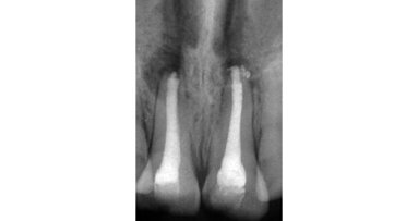
Fig. 1: Initial clinical radiograph showing tooth #21 with a previous root canal treatment, a metal post, a full-coverage coronal restoration and an oblique fracture from the mesial to distal aspect.

Fig. 1: Initial clinical radiograph showing tooth #21 with a previous root canal treatment, a metal post, a full-coverage coronal restoration and an oblique fracture from the mesial to distal aspect.

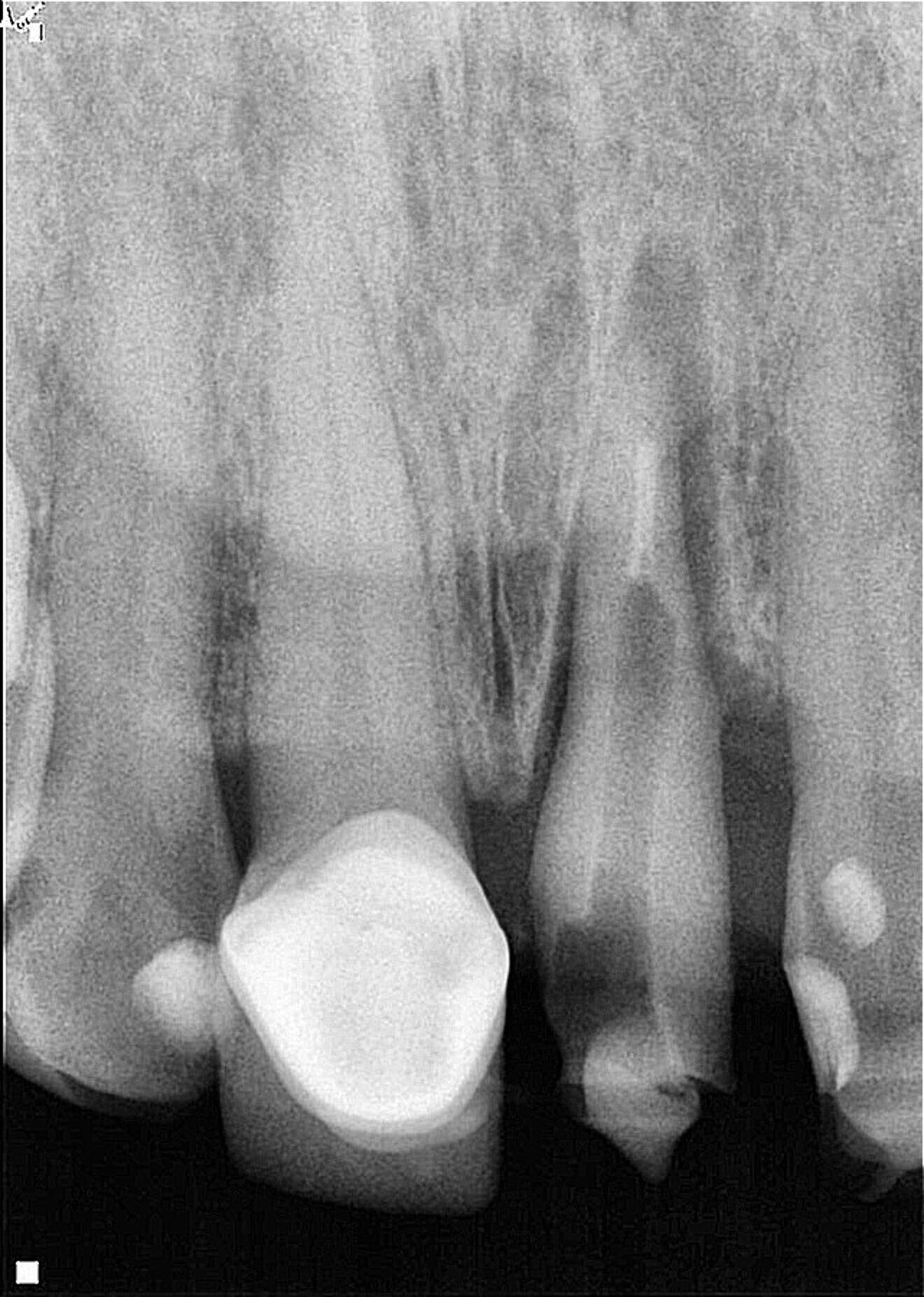
Fig. 2: Post-op radiograph of the tooth extruded by approximately 6 mm and rotated by 180°, locating the deeper zone of the fracture towards the mesial aspect.

Fig. 2: Post-op radiograph of the tooth extruded by approximately 6 mm and rotated by 180°, locating the deeper zone of the fracture towards the mesial aspect.

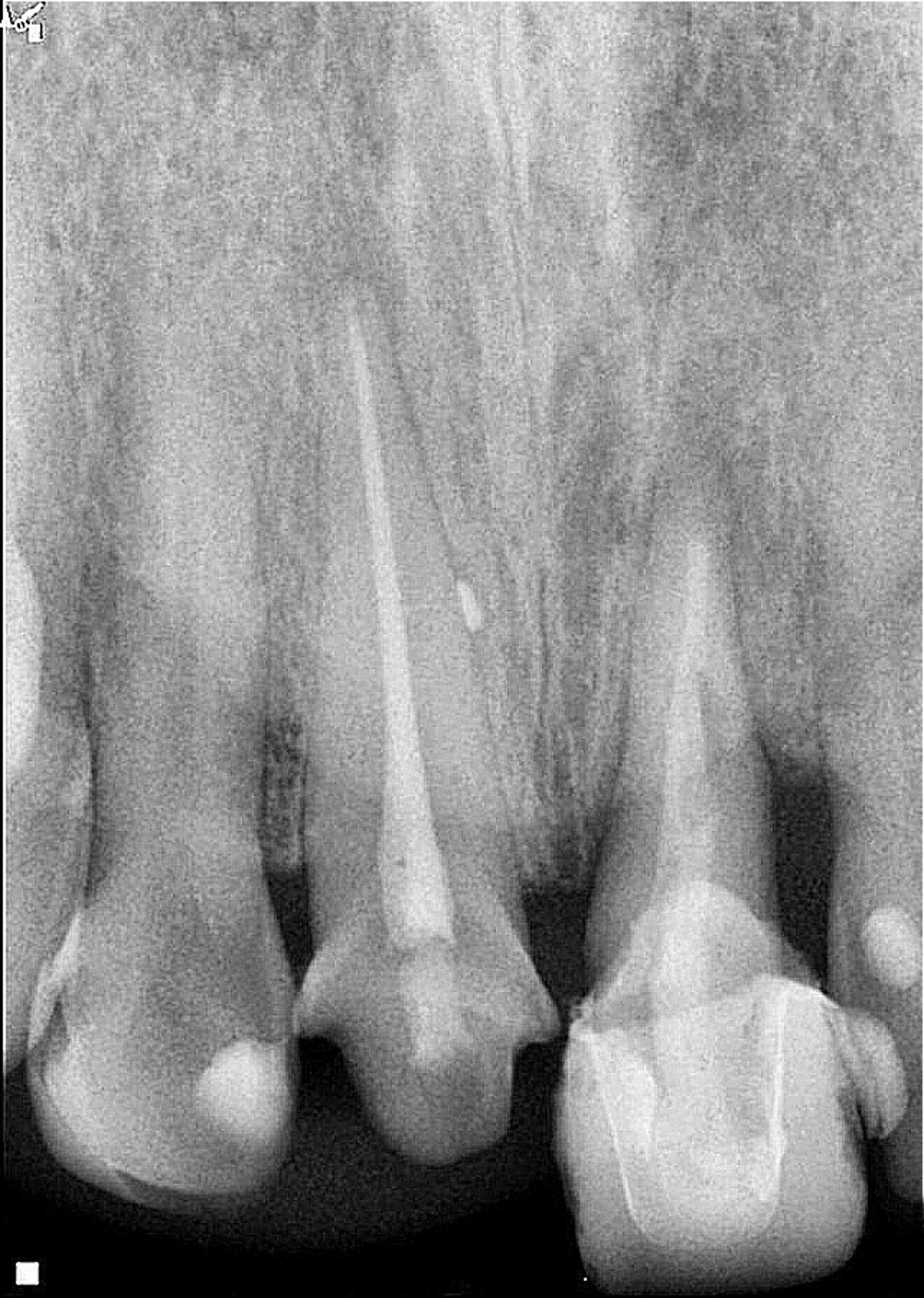
Fig. 3: Eight-week follow-up radiograph with the provisional crown in place showing bone remodelling of the mesial and distal aspects of the bone crest and periapical healing in progress.

Fig. 3: Eight-week follow-up radiograph with the provisional crown in place showing bone remodelling of the mesial and distal aspects of the bone crest and periapical healing in progress.

After removing the metal–porcelain crown and disinfecting the affected area using a gauze dampened with 2% chlorhexidine (GLUCO-CHeX 2%, CERKAMED), syndesmotomy was performed with a No. 15c scalpel, followed by luxation of the root using a small periotome. Luxation forces were applied only to the first 3 mm of the radicular structure at the mesial aspect with the objective of avoiding damage to the periodontal ligament, which was intended to remain in an infra-osseous position. Once the tooth had been loosened, it was extruded by approximately 6 mm and rotated by 180° (Fig. 2). The rotation was performed with the purpose of positioning healthy periodontal ligament fibres in the distal area because the crestal bone already had an oblique defect at this site. Usually the bone follows the extruded periodontal ligament fibres during this type of procedure, making vertical bone gain possible.8, 15, 16 The rotation also allowed us to limit the extent of extrusion. The extrusion and rotation were managed with forceps, grasping only the coronal portion of the tooth, which was planned to remain in a supra-osseous position after the treatment.

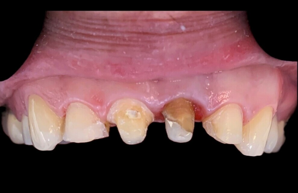
Fig. 4: Clinical situation just before placement of the definitive restoration.

Fig. 4: Clinical situation just before placement of the definitive restoration.

When the tooth reached the desired position, gentle pressure was applied in a labial–palatal direction using a gauze soaked with chlorhexidine to achieve haemostasis before splinting the tooth. While interdental sutures for stabilising the tooth are the first choice according to the literature,17 we chose to use nylon monofilament line with a diameter of 1 mm. This decision was based on its cost-effectiveness, availability, flexibility for splinting, and non-plaque-retentive surface, facilitating good oral hygiene maintenance by the patient. The splint was applied using a flowable composite resin (Nexcomp Flow, Meta Biomed) and a two-step adhesive protocol (EZ Bond Universal, Meta Biomed). The splinting duration was set at three weeks, as recommended to achieve dental stability and minimise the risk of ankylosis.17, 18 The literature indicates that mobility will decrease considerably after a period of two to four weeks after splint removal.6 The patient was provided with oral hygiene instructions and advised to use a soft toothbrush to avoid disturbing the gingival healing.

During a second visit, three weeks after the extrusion, a non-surgical retreatment was performed with the splint in position. At the same appointment, a fibre post was placed to provide adequate core height. The fibre post was cemented using a dual-polymerising resin cement, which was utilised at the same time to rebuild the coronal structure. Before coronal build-up, the splint was removed.

The coronal preparation for the placement of a provisional crown was performed eight weeks after the surgical extrusion procedure. At that appointment, active bone remodelling, mainly at the periapical area, was observed (Fig. 3). This remodelling process is characterised by bone loss at the crestal area in the initial six to eight weeks postoperatively.16 The definitive restoration was placed three months after the initial procedure to provide enough time for the periodontal tissue to heal, thereby preventing post-restorative gingival migration.19, 2 Healthy periodontal tissue, good symmetry of the gingival margin and appropriate ferrule length were observed just before placement of the definitive restoration (Fig. 4).

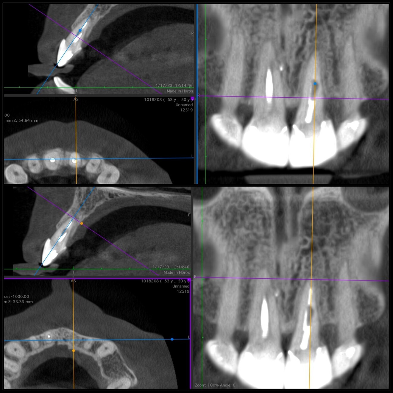
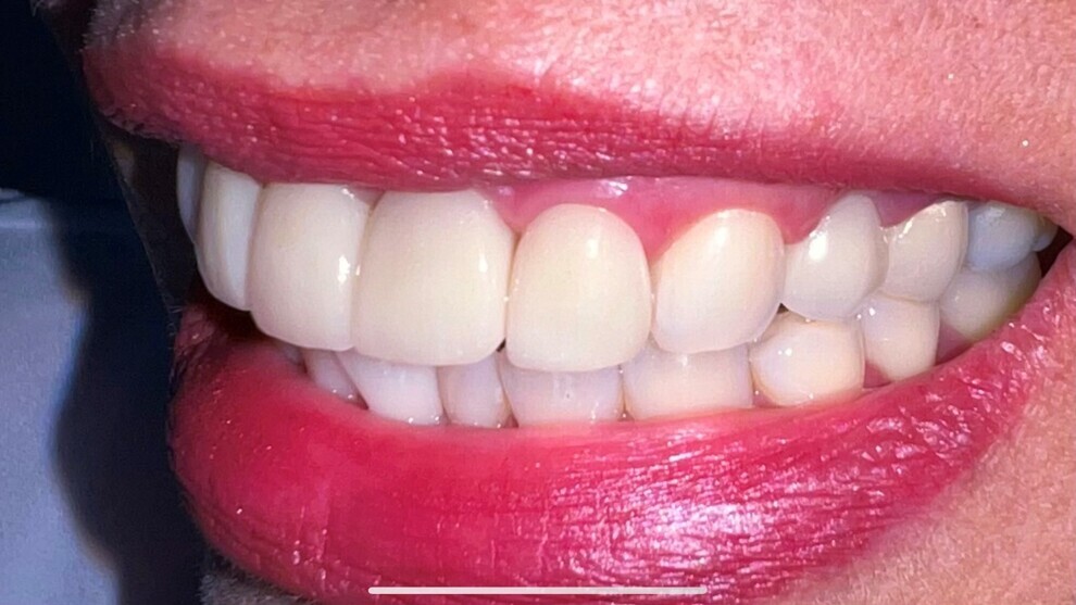
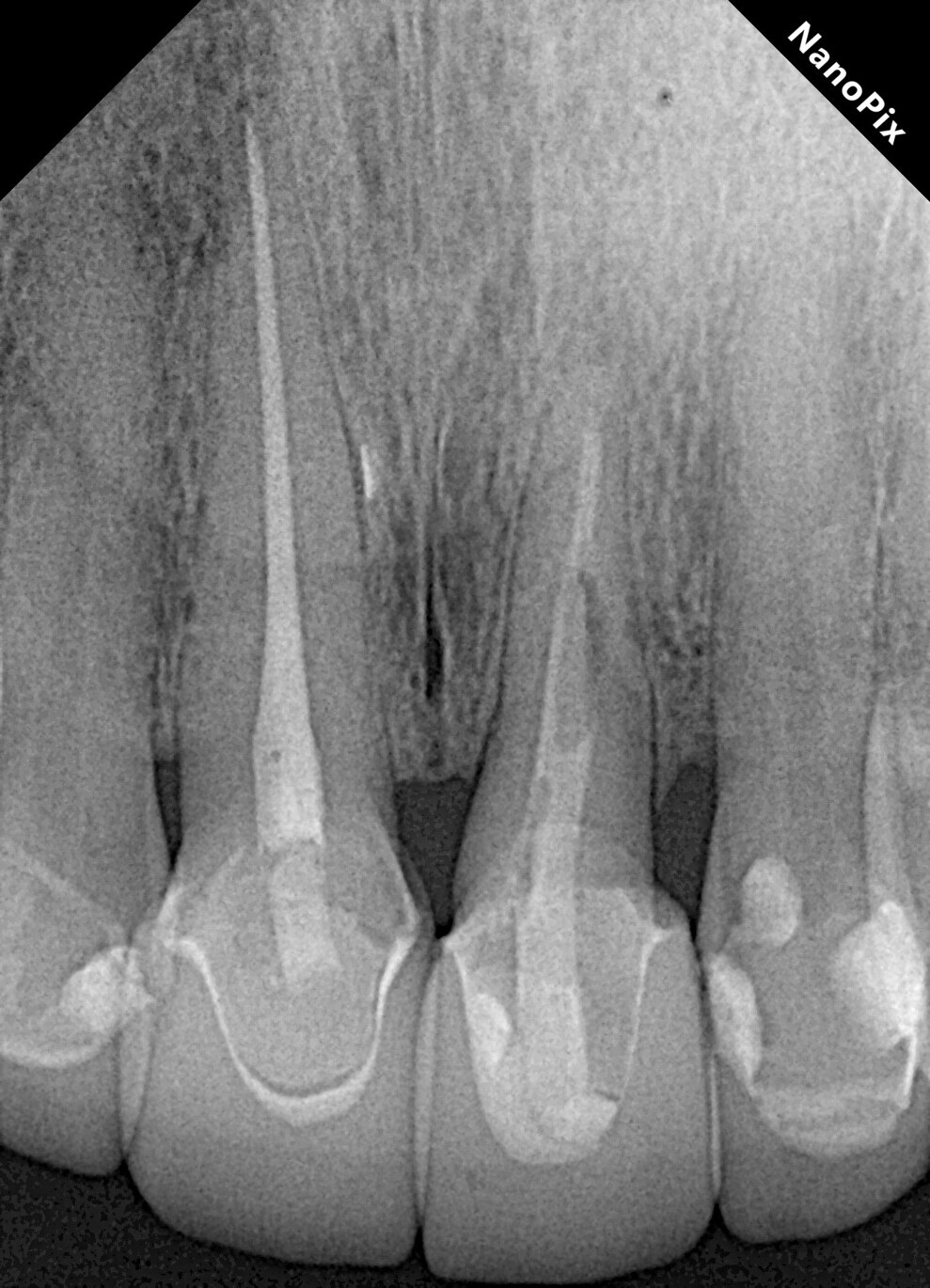
Radiographic examination conducted two years after the extrusion revealed good periapical healing and ongoing vertical bone growth at the interdental bone crest, mainly in the distal area (Fig. 5). It has been reported that a normal periodontal ligament contour may be observed three months postoperatively,6, 8, 20 and periapical repair and resolution of radiolucency are commonly seen six months later, as well as minimal marginal bone loss,6 often associated with bone damage incurred during the extrusion process. After two years of follow-up, complete repair of the periapical tissue and ongoing bone remodelling mainly in the area of the distal bone crest were observed (Fig. 6). The tooth remains asymptomatic, non-mobile, and fully functional aesthetically and biologically (Fig. 7). The radiographic situation after the four-year follow-up period showed stable results (Fig. 8).

Fig. 5: Two-year follow-up radiograph showing periapical healing and signs of bone remodelling, mainly vertical bone growth in the distal area.

Fig. 5: Two-year follow-up radiograph showing periapical healing and signs of bone remodelling, mainly vertical bone growth in the distal area.

Fig 6: Two-year follow-up CBCT scan showing complete periapical healing, bone remodelling in the distal area and no signs of resorption.

Fig 6: Two-year follow-up CBCT scan showing complete periapical healing, bone remodelling in the distal area and no signs of resorption.

Discussion

The basic principle of biological width preservation relies on avoiding its invasion during restorative procedures.21, 22 When crown lengthening is planned to increase supragingival coronal length, anatomical and biological considerations are necessary. Different techniques for crown lengthening procedures have been proposed, particularly in the aesthetic zone, where preserving the gingival margin and interdental papillae is essential for achieving satisfactory aesthetic outcomes.19, 21, 23

Fig. 7: Clinical photograph showing good adaptation of the coronal restoration, healthy periodontal tissue and a good aesthetic result.

Fig. 7: Clinical photograph showing good adaptation of the coronal restoration, healthy periodontal tissue and a good aesthetic result.

Surgical extrusion aims to separate the periodontal ligament using surgical instruments such as fine elevators, periotomes, forceps and scalpels to reposition the tooth in a more coronal position and thereby obtain a healthier supragingival structure for an adequate definitive restoration, without risking damage to the periodontal tissue and invasion of the biological width.8, 17, 24 This procedure is indicated in cases of corono-radicular fracture,16, 25 root fracture,26 carious lesions extending below the alveolar crest,27 cervical root resorption and insufficient supragingival dental structure to provide an adequate ferrule effect.16, 17, 28 In cases of subgingival labial fracture, tooth rotation by 180° may also be indicated to reduce the amount of tooth requiring extrusion.6, 17 In this case, the rotation was also performed with the aim of having healthy periodontal ligament cells in the area of the periodontal oblique defect to increase the possibility of vertical bone gain.15 In some cases, it is important to limit the amount of extrusion to maintain an adequate crown–root ratio, and rotation can also be beneficial in this regard.

Some surgical extrusion techniques reported in the literature involve flap elevation with careful apex exposure, but these techniques might compromise the aesthetics in the anterior; more conservative approaches are increasingly favoured.6 Studies have reported that the use of axial traction forces significantly reduces cementoblast loss on root surfaces compared with tooth extraction with forceps.29 One disadvantage of the technique suggested in this report for luxating the tooth is that, if the clinician is not gentle when applying pressure, there is a possibility of exerting high compressive forces on the apical and corono-radicular periodontal ligament. This can cause damage to cementoblasts and periodontal ligament cells at some points around the root surface, consequently increasing the risk of superficial root resorption.29, 30 However, superficial root resorption in these cases is not progressive in nature and is primarily a transient consequence of the surgery.16, 31 Higher rates of root resorption have been observed in cases where root canal treatment was performed before the extrusion procedure.6
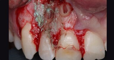
Although bone grafting has been reported to improve tissue repair at the periapical level,11, 24 its benefits have not been clearly demonstrated. However, periapical tissue disruption leads to the formation of an apical clot, which matures into fibrous scaffolding and subsequently into cancellous bone;32, 33 for this reason, bone grafting was not considered a treatment option in this case.

Fig. 8: Four-year follow-up periapical radiograph showing absence of inflammatory signs in the periapical area, absence of signs of root resorption, bone gain in the area of the distal bone crest and a normal and healthy periodontal ligament space.
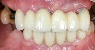
Fig. 8: Four-year follow-up periapical radiograph showing absence of inflammatory signs in the periapical area, absence of signs of root resorption, bone gain in the area of the distal bone crest and a normal and healthy periodontal ligament space.

Some advantages of the surgical extrusion technique include shorter operative and overall restoration times compared with orthodontic extrusion, the relative ease of performing the procedure, lower cost compared with conventional crown lengthening techniques, brief consultation time, uncomplicated recovery and minimal long-term patient cooperation. Possible adverse events associated with the technique presented in this case report may include root fracture during the extrusion procedure, superficial root resorption, marginal bone loss, persistent dental mobility, tooth loss due to compromise of the periodontal ligament or an inadequate crown–root ratio, and dental ankylosis (not common with this technique because all procedures are performed under controlled surgical conditions).1–3, 8, 16, 17 Other limitations related to this procedure may include patient acceptance, difficulty in aesthetic provisionalisation of the tooth, establishing good oral hygiene in the postoperative period (mainly during the first postoperative week) and controlling bleeding when placing the flexible adhesive splint.8, 17

Conclusion

The four-year follow-up of the surgical extrusion case presented in this report showed absence of inflammatory signs at the periapical area, absence of signs of root resorption, bone gain in the area of the distal bone crest, and a normal and healthy periodontal ligament space. Clinically, the tooth remains fully functional aesthetically and biologically, demonstrating a satisfactory long-term outcome of the clinical technique presented.

Conflict of interest

The authors have no conflicts of interest to declare.

Editorial note:

The complete list of references can be found here.

Topics:
Tags:
To post a reply please login or register
advertisement
advertisement