Initial situation
A 38-year-old female patient presented to the dental office reporting social discomfort when smiling, owing to the absence of both maxillary second premolars (Figs. 1a– c, Figs. 2a–c & 3). Clinical examination revealed that tooth #16 had an intra-radicular post and had been prepared for a crown, but the crown was missing, and a cavity was noted on the distal surface of tooth #14. Overall, the patient exhibited a healthy periodontal condition. She was a non-smoker and did not present with any comorbidities or systemic health issues. CBCT imaging showed adequate bone quality and quantity at both sites and no signs of acute infection. Additionally, a residual root was identified at site #15 beneath the already healed gingiva.
Treatment planning
The patient’s maxillary and mandibular arches were scanned using the Straumann SIRIOS system. The scans, along with the DICOM files from the CBCT scan, were sent via Straumann AXS to Smile in a Box (Straumann) for treatment planning, surgical guide design and 3D printing of the model and surgical guides (Figs. 4, 5a–f & Figs. 6a–d).
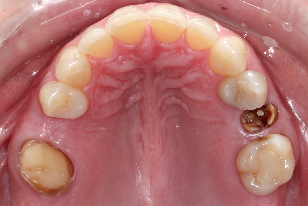
Fig. 3: Occlusal view of the initial situation.
After evaluation and validation of the plan, it was decided for site #15—where a residual root was present and the bone density was generally soft—that a Straumann BLX (Roxolid, SLActive, regular base) 4.5 × 8.0 mm implant would be placed, along with an M shape Straumann Anatomic Healing Abutment XC (regular base/wide base; gingival height: 1.5 mm). For site #25, extraction of the remaining root was planned and would be followed by the placement of a Straumann BLC (Roxolid, SLActive, regular base) 3.75 × 8.00 mm implant. This narrower site had a visible lamina dura, favourable for achieving primary stability. An M shape Straumann Anatomic Healing Abutment XC (regular base/wide base; gingival height: 1.5 mm) was also selected for this site to support proper soft-tissue emergence during the 60- to 90-day healing period. Owing to the expected post-extraction gaps between the implants and buccal bone walls, cerabone plus (botiss biomaterials) was planned to be used for grafting.
After the healing and gingival maturation phases, the patient would return for intra-oral scanning directly on the healing abutments, eliminating the need for abutment removal thanks to their scannable head design. Definitive restorations were planned as full-contour monolithic zirconia crowns cemented on to Straumann Variobase abutments.
Surgical procedure
Under local anaesthesia administered bilaterally, the surgical procedure began at site #15 with a supra-crestal incision and a full-thickness flap to ensure full visibility of the area for the location and extraction of the residual root previously identified on the CBCT scan. The root fragment was located and extracted without complication. At site #25, a minimally traumatic, flapless extraction was performed using a periotome, followed by the use of forceps to gently remove the root (Figs. 7a–h).
The surgical guide was inserted and verified for accurate fit. Osteotomy preparation was carried out according to the protocol generated by coDiagnostiX (Dental Wings), tailored to the planned implant types on each side (Figs. 8a–g). The Straumann BLX implant was placed into site #15, achieving primary stability at 30 N cm. The Straumann BLC implant was inserted into site #25, reaching primary stability at 50 N cm (Figs. 9a–g).
To preserve the ridge contour over time, cerabone plus was hydrated according to the manufacturer’s instructions and placed into the buccal gap at site #25 (Figs. 10a–f). Both sites received healing abutments and were sutured using mesial and distal single interrupted sutures to ensure soft-tissue stability during the healing phase (Figs. 11a–f & Figs. 12a–d). The patient received postoperative instructions along with an analgesic prescription and was scheduled for suture removal after two weeks.
Prosthetic procedure
After 60 days, the patient returned to the clinic for intra-oral scanning to initiate the definitive crown workflow. A noteworthy advantage of the Straumann Anatomic Healing Abutment XC is that it allows direct scanning without removal, eliminating the need for a scan body. This significantly minimises soft-tissue disturbance, enhances patient comfort and reduces overall chair time.
Intra-oral scanning was performed using Straumann SIRIOS (Fig. 13), and the files were exported to CARES (Straumann) for definitive crown design (Fig. 14). The virtual subgingival contour provided by the Straumann Anatomic Healing Abutment XC in the software can be easily replicated in the definitive restoration, offering a substantial prosthetic benefit.
Two monolithic zirconia crowns were milled and cemented on to Straumann Variobase abutments with the same gingival height as that of the healing abutments. The crowns were tested on a 3D-printed model to evaluate occlusion and excursive movements (Figs. 15a–c). Additionally, site #16 received a temporary three-quarter onlay.
Once the definitive crowns were ready, the healing abutments were removed, revealing a natural gingival contour—ideal for receiving the definitive restorations without discomfort or disruption to the peri-implant tissue (Figs. 16a–c, 17a & b). Both crowns were seated, and the retaining screws were torqued to 35 N cm, as recommended for Straumann final abutments. The screw channels were protected with PTFE tape and sealed with flowable composite. Occlusion and excursive movements were carefully checked, and the patient received comprehensive hygiene instructions (Figs. 18a–c).
Treatment outcomes
The treatment process and outcomes exceeded the patient’s expectations, in that the total treatment time was only 65 days and she experienced no discomfort throughout the process. At the one-year follow-up appointment, new photographs and radiographs were taken, revealing healthy and stable soft and hard tissue (Figs. 19a–d).

 Brazil / Brasil
 Canada / Canada
 Latin America / Latinoamérica
 USA / USA
 Austria / Österreich
 Bosnia and Herzegovina / Босна и Херцеговина
 Bulgaria / България
 Croatia / Hrvatska
 Czech Republic & Slovakia / Česká republika & Slovensko
 France / France
 Germany / Deutschland
 Greece / ΕΛΛΑΔΑ
 Hungary / Hungary
 Italy / Italia
 Netherlands / Nederland
 Nordic / Nordic
 Poland / Polska
 Portugal / Portugal
 Romania & Moldova / România & Moldova
 Slovenia / Slovenija
 Serbia & Montenegro / Србија и Црна Гора
 Spain / España
 Switzerland / Schweiz
 Turkey / Türkiye
 UK & Ireland / UK & Ireland
 China / 中国
 India / भारत गणराज्य
 Pakistan / Pākistān
 Vietnam / Việt Nam
 ASEAN / ASEAN
 Israel / מְדִינַת יִשְׂרָאֵל
 Algeria, Morocco & Tunisia / الجزائر والمغرب وتونس
 Middle East / Middle East

 Dr. Paul Renton HarperRegister now1CELive webinar

 Assoc. Prof. Aaron Davis, Prof. Sarah BakerRegister now1CELive webinar
 Dr. Vasiliki Maseli DDS, MS, EdMRegister now1CELive webinar
 MDT Andreas ChatzimpatzakisRegister now1CE


, and flapless extraction in site #25 (e–h).")

, and Straumann BLC implant placed in site #25 (e–g).")
 for gap filling.")

 and radiographic situation two weeks after surgery (c & d).")


 and 25 after abutment removal (b).")


 and occlusal views showing stable soft- and hard-tissue levels at the one-year follow-up (c & d).")


To post a reply please login or register