The procedure of implantation is becoming an increasingly popular method for replacing teeth. The critical factor in the achievement of a therapeutic and aesthetic long-term effect is the accuracy and precision of implant placement, being the support for the future prosthetic work.
Thanks to modern digital technologies, it is possible to plan the implantation virtually. Evaluation of this plan by 3D printing in a subsequent step allows the creation of implant guides. Using the guides, which provide precise information on implant placement and insertion depth and angle, allows the maintenance of all the parameters included in the planning stage, lowering the risk of a mistake during implantation. Using 3D printing allows the fabrication of both implant guides and study models that accurately represent patients’ true clinical conditions. This makes it possible to compare the precision of procedures under the in vitro conditions, which are safe and representative of actual requirements. During the implantation, clinical conditions very often hinder precise orientation in the operating field, thus the precision of implant positioning is lower. According to the literature, both more and less experienced clinicians face this problem. Introducing virtual planning based on CBCT is highly useful while preparing for the procedure; however, what allows the fully controlled preparation of the implantation site is the transfer of its result to the guide imposing the positioning. The virtually created implant guide can be printed using a 3D printer, sterilised and then used in the procedure. The use of the guide affects the precision of the procedure and shortens its time.
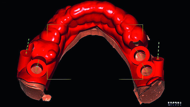
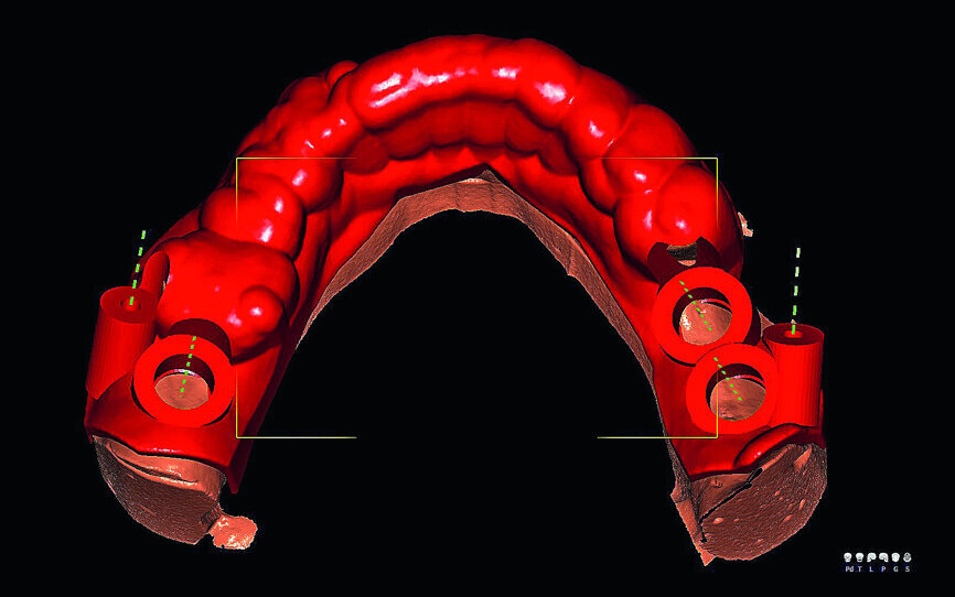
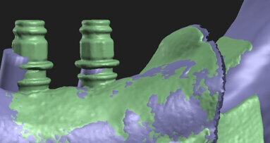
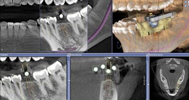
Fig. 1a: Virtual planning of implant positioning.
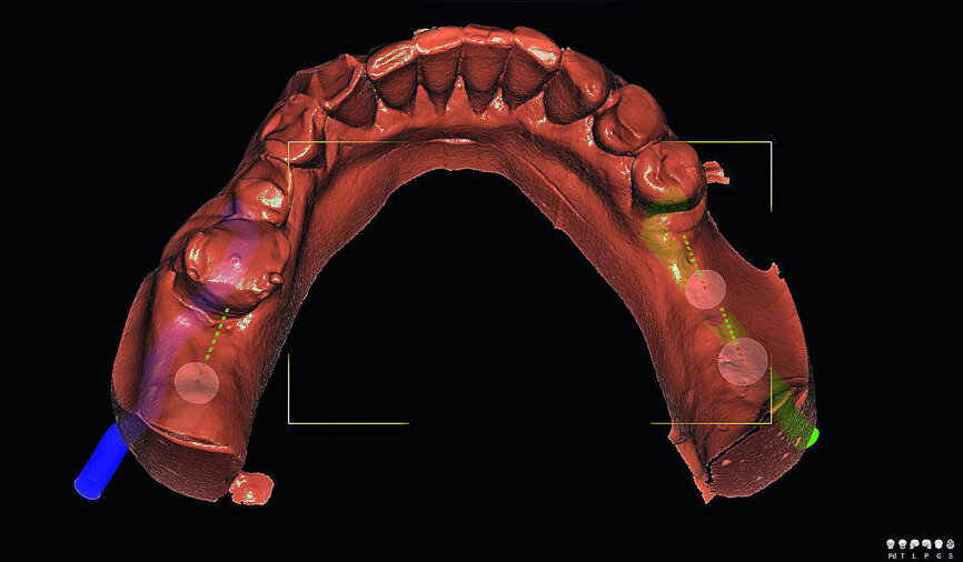
Fig. 1b: Virtual planning of implant positioning.
Fig. 2: Exemplary pair of models before the procedure: on the left, without the guide, and on the right, with the guide.
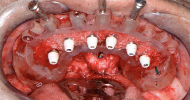
Fig. 3: A model with the guide and implants after the implantation. The guide was stabilised with two posts.
Fig. 4: The material was deposited on the drill attached to the extension.
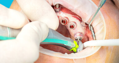
Fig. 5: Drill attached to the extension passing through the reduction sleeve. The extension allows the drill to be guided correctly without touching the template on the adjacent tooth with the contra-angle handpiece. Clinically, the use of the drill extension may be impossible, especially in molars, owing to the limited opening of the jaws.
Fig. 6: Models after performing the procedures with the use of the guide. The same of the implants inserted are visible.
Aim of the study
The aim of the study is to prepare 3D models for the analysis of the precision of implant procedures performed on the basis of digital planning, conducted with and without the use of implant guides.
Methodology
Based on the CBCT examination of the patient, who underwent implantation in the mandible, a 3D model corresponding to the actual bone and mucosal conditions before implantation was created in DDS-Pro software (www.dds-pro.com.pl). It was then reprinted 20 times. The print was produced with selective laser sintering technology using polyamide powder in the TPM Elite P3600 SLS System printer (Solveere). It yielded ten identical pairs of mandibular models. Virtual planning ( DDS-Pro; Fig. 1) of implant positioning and placement (TSIII, OSSTEM IMPLANT) and the implant guide, printed in 3D with Jet technology (ProJet MP 3000 printer, 3D Systems), with stock sleeves for three implants with regular platforms previously used clinically (sterilised), were used to introduce implants into every second printed model, using the OsstemGuide KIT(Taper). The drilling speed was set at 1,200 rpm. Water cooling was not used. Osteotomies were performed according to the manufacturer’s instructions. Other models were used for implantation based on the planning performed, but without additional help (no guide), using the same implant kit and under the same conditions. As the test was conducted in vitro, TSIII training implants with dimensions of 4 × 10 mm were used. It was assumed that all ten procedures performed would yield the same results.
Findings
The use of 3D printed models allows implantation under conditions spatially corresponding to those of a clinical situation. However, the models printed in this study were hard. The material cut during osteotomy preparation was deposited on the drill and the implant thread, making it difficult to perform full-depth insertion. More torque was required to insert the implant than is clinically used. It was observed that, when an osteotomy was prepared in the vicinity of a preserved tooth, there was a need to use the drill extension in order to avoid leaning the contra-angle handpiece on the guide or tooth. Because this tool is missing in the OsstemGuide KIT(Taper), one must have an additional implant kit when using it clinically. The use of the guide shortens the implantation time, compared with the same procedure performed with no help of a guide.
In the following stage of the project, the models will be optically scanned and undergo comparative analysis in terms of repeatability, accuracy and compliance with the planned virtual goal.
Editorial note: The study is being carried out as a part of a project in the field of scientific developmental research aimed at the development of young scientists and students enrolled in PhD studies, financed as part of the scientific activity of the Medical University of Warsaw in Poland. This article was published in CAD/CAM - international magazine of digital dentistry No. 01/2019.
Proper 3D implant positioning is a crucial factor for predictable implant and prosthodontic treatment. Guided osteotomy preparation and placing implants ...
When a patient presents to your dental practice with questionable and/or non-restorable teeth requiring full-mouth extractions, the greatest concern is ...
The evolution of digital dentistry has revolutionised the way dental procedures are planned and executed. This case highlights the integration of various ...
When a patient presents to your dental practice with questionable and/or non-restorable teeth requiring full-mouth extractions, the greatest concern is ...
Implant placement in spaces that are narrow in the mesiodistal dimension poses challenges related to the surgical aspect of treatment. This is further ...
BERLIN, Germany: According to implantology and periodontics specialist Dr Juan Arias Romero, a new synergy between digital planning, guided implant ...
In cases of severe posterior bone atrophy, Straumann Pro Arch is a solution that helps achieve fixed restoration for the patient. Straumann Guided Surgery ...
A 62-year-old male patient was referred to my practice for implant planning and treatment in the maxillary anterior region. The teeth in the maxillary ...
The objective of this review was to evaluate the scientific evidence on accuracy, as well as esthetic and clinical outcomes of single-tooth implants placed ...
Guided surgery has been around for a long time. However, very few dentists in the UK place implants using surgical guides. The reasons for this are ...
Live webinar
Wed. 25 February 2026
11:00 am EST (New York)
Prof. Dr. Daniel Edelhoff
Live webinar
Wed. 25 February 2026
1:00 pm EST (New York)
Live webinar
Wed. 25 February 2026
8:00 pm EST (New York)
Live webinar
Tue. 3 March 2026
11:00 am EST (New York)
Dr. Omar Lugo Cirujano Maxilofacial
Live webinar
Tue. 3 March 2026
8:00 pm EST (New York)
Dr. Vasiliki Maseli DDS, MS, EdM
Live webinar
Wed. 4 March 2026
12:00 pm EST (New York)
Munther Sulieman LDS RCS (Eng) BDS (Lond) MSc PhD
Live webinar
Wed. 4 March 2026
1:00 pm EST (New York)


 Austria / Österreich
 Bosnia and Herzegovina / Босна и Херцеговина
 Bulgaria / България
 Croatia / Hrvatska
 Czech Republic & Slovakia / Česká republika & Slovensko
 France / France
 Germany / Deutschland
 Greece / ΕΛΛΑΔΑ
 Hungary / Hungary
 Italy / Italia
 Netherlands / Nederland
 Nordic / Nordic
 Poland / Polska
 Portugal / Portugal
 Romania & Moldova / România & Moldova
 Slovenia / Slovenija
 Serbia & Montenegro / Србија и Црна Гора
 Spain / España
 Switzerland / Schweiz
 Turkey / Türkiye
 UK & Ireland / UK & Ireland
 Brazil / Brasil
 Canada / Canada
 Latin America / Latinoamérica
 USA / USA
 China / 中国
 India / भारत गणराज्य
 Pakistan / Pākistān
 Vietnam / Việt Nam
 ASEAN / ASEAN
 Israel / מְדִינַת יִשְׂרָאֵל
 Algeria, Morocco & Tunisia / الجزائر والمغرب وتونس
 Middle East / Middle East














To post a reply please login or register