Search Dental Tribune

This clinical case describes a full-arch rehabilitation combining dynamic navigation for maxillary zygomatic implants, task-autonomous robotics for mandibular implant placement and intra-oral photogrammetry for immediate provisionalisation. (All images: Drs James Chow & Nikos Mattheos)

Tue. 15. September 2026

save

Computer-assisted implant surgery (CAIS) is no longer a single technology but a workflow centred on the digital treatment plan and involving several distinct treatment execution modalities—static guides, dynamic navigation and task-autonomous robotics—each with its own accuracy profile, training requirements and anatomical suitability. 1, 2 A recent international white paper on the clinical outcomes of CAIS concluded that all guided modalities improve implant placement accuracy compared with free-hand surgery, but that the clinically meaningful benefit lies in matching the modality to the complexity of the individual surgical task and the patient’s needs, rather than defaulting to a single CAIS modality throughout a treatment plan.1 The challenge for clinicians is therefore to orchestrate an efficient workflow using a wide range of software and hardware systems that are often incompatible.3

Zygomatic implant placement, particularly the quad zygoma protocol, in which four implants support a fixed prosthesis in a severely atrophic edentulous maxilla, remains one of the most demanding applications of guided implant surgery. This is due to the long, angulated trajectories and the proximity of the orbit, skull base and nasal cavity.4 Dynamic navigation is currently the most extensively documented guided modality for this application, and prospective data support its accuracy in full-arch zygomatic and conventional implant placement for immediate loading.4, 5 Task-autonomous robotic placement of zygomatic implants has also been described, but the evidence is at an early stage and is largely from in vitro and ex-vivo studies, and case reports. A recent systematic review highlighted the limited evidence base compared with that for conventional implant placement.6

By contrast, a growing body of clinical literature has evaluated the accuracy and effectiveness of task-autonomous robotic systems for conventional implant placement.7 Yakebot (Beijing Yakebot Technology), a task-autonomous system that executes the planned osteotomy and implant placement under continuous operator supervision, has been reported to achieve significantly smaller platform, apical, depth and angular deviations compared with fully guided static CAIS in edentulous jaws.8 A recent meta-analysis of robotic CAIS trueness across clinical studies reached similar conclusions.9

Recording the spatial relationship of four widely divergent zygomatic implant positions across a full arch presents a prosthetic registration challenge that can be difficult to manage with conventional impression techniques, particularly when immediate provisionalisation is required. In a recent prospective clinical study, intra-oral photogrammetry enabled accurate full-arch capture of implant positions and immediate loading of a 3D-printed provisional prosthesis.10

This report describes a comprehensive full-mouth rehabilitation in which two CAIS modalities together with photogrammetry were combined: dynamic navigation for zygomatic implant placement in the maxilla, task-autonomous robotics for conventional implant placement in the mandible and intra-oral photogrammetry for fullarch immediate provisionalisation. Each CAIS technology was matched to the anatomical characteristics of the arch in which it would be used, rather than applying one CAIS modality uniformly.

Case presentation

A 59-year-old male patient, who smoked approximately five to ten cigarettes per day, presented with a heavily restored and failing dentition in both arches. His medical history included hypertension and hyperlipidaemia, managed with amlodipine and simvastatin, respectively. He also reported taking omega-3, co-enzyme Q10 and glucosamine supplements. His body mass index was 28.6 kg/m² (78 kg; 1.65 m).

Clinical examination revealed bimaxillary protrusion, loss of posterior support, and anterior teeth compromised by recurrent caries and defective metal–ceramic and cast restorations (Figs. 1a & b). Panoramic radiography and CBCT demonstrated extensive bilateral pneumatization of the maxillary sinuses, extending anteriorly to the central incisor region. A 3D virtual patient was created for diagnosis and treatment planning using data from CBCT, intra-oral and facial scanning and dynamic jaw motion registration (SHINING 3D).


Fig. 1a: Preoperative situation. Extra-oral frontal smile view showing the heavily restored and severely compromised dentition, including recurrent caries and defective restoration margins.

Fig. 1a: Preoperative situation. Extra-oral frontal smile view showing the heavily restored and severely compromised dentition, including recurrent caries and defective restoration margins.

Fig. 1b: Intra-oral frontal view with the teeth in maximum intercuspation, showing extensive tooth wear and multiple failing restorations.

Fig. 1b: Intra-oral frontal view with the teeth in maximum intercuspation, showing extensive tooth wear and multiple failing restorations.

Diagnosis

The patient was diagnosed with terminal dentition in both arches. The severity of atrophy and bilateral maxillary sinus pneumatisation precluded conventional implant rehabilitation, including the use of short or tilted implants, and the anatomy was considered unsuitable for trans-nasal or pterygoid implant anchorage (Figs. 2a & b). This anatomical assessment indicated a quad zygoma approach in the maxilla, combined with conventional implant-supported rehabilitation in the mandible after extraction of the remaining dentition in both arches.

Treatment plan and implant details

The treatment plan comprised: (1) extraction of the remaining maxillary and mandibular dentition; (2) placement of four zygomatic implants (NobelZygoma 45°, external hex, TiUltra surface; Nobel Biocare) in the maxilla using dynamic computer-assisted navigation (X-Guide, X-Nav Technologies), followed by the use of intra-oral photogrammetry (FastMap, X-Nav Technologies) to capture the spatial relationship of the four multi-unit abutment positions for immediate provisionalisation; and (3) placement of four endosseous implants in the mandible (Straumann BLX, Roxolid) using the Yakebot robotic system (Figs. 3a & b).

The rationale for this arch-specific approach reflected the different demands of each arch. The long, angulated trajectories of the zygomatic implants and their proximity to critical structures favoured a mature and well-documented dynamic navigation platform, offering full guidance but still allowing full free-hand control by the surgeon.

The shorter, more standardised mandibular osteotomies were considered for task-autonomous robotic execution, for which clinical evidence on placement accuracy is more extensive. The virtual patient was used to demonstrate the proposed treatment and discuss alternatives with the patient. Given the complexity of the rehabilitation, the extent and cost of treatment were also discussed with the patient and his family.

Surgical protocol and timeline

The two surgical procedures were performed 11 days apart, and the maxillary provisional prosthesis was delivered on the fourth day after maxillary surgery and the mandibular provisional prosthesis on the third day after mandibular surgery. The dynamic navigation and robotic CAIS workflows described in a recent publication were followed.3

Maxillary surgery was performed under general anaesthesia, and local anaesthetic was administered by infiltration. A full-thickness flap was raised, followed by resection of the anterior maxilla with a piezoelectric saw and removal of the remaining maxillary root remnants (teeth #14–24) and associated apical granulomas. In addition, two non-restorable mandibular left molars (teeth #36 and 37) were extracted. Piezoelectric surgery was used for bone recontouring. A rigid optical reference tracker was fixed to the maxilla with two vertically positioned bone screws.

For the dynamic navigation system, the patient’s anatomy was then registered to the digital treatment plan using the X-Mark protocol, and four zygomatic implants were placed through the body of the zygomatic bone and multi-unit abutments attached (Fig. 4):

  • A 4.0 × 45.0 mm implant was placed in site #16 to an insertion torque of > 40 Ncm, followed by attachment of a short abutment (collar height: 3 mm) tightened to 35 Ncm.
  • A 4.0 × 60.0 mm implant was placed in site #12 to an insertion torque of > 60 Ncm, followed by attachment of a short abutment (collar height: 3 mm) tightened to 35 Ncm.
  • A 4.0 × 60.0 mm implant was placed in site #22 to an insertion torque of > 60 Ncm, followed by attachment of a medium abutment (collar height: 5 mm) tightened to 35 Ncm.
  • A 4.0 × 42.5 mm implant was placed in site #26 to an insertion torque of > 50 Ncm, followed by attachment of a medium abutment (collar height: 5 mm) tightened to 35 Ncm.
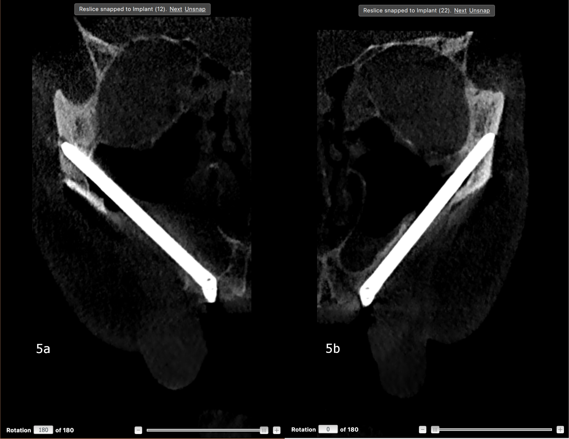
Figs. 5a & b: Postoperative CBCT slices showing the zygomatic implants emerging at sites #12 (a) and 22 (b).

Figs. 5a & b: Postoperative CBCT slices showing the zygomatic implants emerging at sites #12 (a) and 22 (b).


A collagenated xenogeneic bone substitute (three 250 mg units) and a resorbable collagen membrane (30 × 40 mm) were placed at the extraction and augmentation sites. The flap was closed with #4-0 Vicryl sutures and a postoperative CBCT scan was obtained to confirm the implant positions (Figs. 5a & b). Intra-oral photogrammetry was used to capture the spatial relationship of the four multi-unit abutment positions directly, allowing fabrication of a provisional fixed prosthesis without a conventional multi-unit abutment impression (Fig. 6).

Four days later, the maxillary provisional prosthesis was delivered (Fig. 7). The occlusion was adjusted, the fit was confirmed radiographically and a night guard was provided.

Fig. 6: FastMap photogrammetry interface showing registration of the four scan bodies connected to the zygomatic implants.

Fig. 6: FastMap photogrammetry interface showing registration of the four scan bodies connected to the zygomatic implants.

Fig. 7: Intra-oral frontal view of the maxillary provisional prosthesis delivered on the fourth day after maxillary surgery.

Fig. 7: Intra-oral frontal view of the maxillary provisional prosthesis delivered on the fourth day after maxillary surgery.

Eleven days after maxillary surgery, mandibular surgery was performed under general anaesthesia, and local anaesthetic was administered by infiltration. Four bone-anchored fiducial markers were placed using a surgical guide for registration of the mandible with the robotic system (Figs. 8a &b). The remaining mandibular dentition was then extracted. Flapless osteotomy preparation and implant placement were performed autonomously by the robotic arm under continuous surgeon supervision, based on the drilling plan and real-time tracking data (Figs. 9–12), and straight abutments were attached:

A minimally invasive flap was raised, and piezoelectric surgery was used for bone reduction to establish a flat prosthetic platform. The flap was closed with #4-0 Vicryl sutures, and a postoperative CBCT scan was obtained. An implant-level digital impression was then recorded using intra-oral photogrammetry (Aoralscan Elite 4D, SHINING 3D; Fig. 13).

Three days later, the mandibular provisional prosthesis was delivered (Figs. 14a & b). The occlusion was checked, passive fit was confirmed radiographically and continued use of the night guard was reinforced.

Definitive restorative phase

At 3.5 months postoperatively, a dataset for both arches was acquired using intra-oral and facial scanning, jaw motion registration and intra-oral photogrammetry (Aoralscan Elite; Figs. 15 & 16). The jaw motion records were transferred to a virtual articulator, and photogrammetric scan body data was recorded for the mandibular and maxillary implants (Figs. 17a & b).

Fig. 13: Photogrammetric scan bodies connected to the mandibular implants immediately after surgery.

Fig. 13: Photogrammetric scan bodies connected to the mandibular implants immediately after surgery.

Fig. 14a: Mandibular provisional prosthesis before delivery.

Fig. 14a: Mandibular provisional prosthesis before delivery.

Fig. 14b: Mandibular provisional prosthesis in occlusion with the maxillary provisional prosthesis after delivery on the third day after mandibular surgery.

Fig. 14b: Mandibular provisional prosthesis in occlusion with the maxillary provisional prosthesis after delivery on the third day after mandibular surgery.

Fig. 15: Intra-oral scan of the provisional prostheses in function for 3.5 months.

Fig. 15: Intra-oral scan of the provisional prostheses in function for 3.5 months.

Fig. 16: Jaw motion registration with the maxillary and mandibular provisional prostheses in place.

Fig. 16: Jaw motion registration with the maxillary and mandibular provisional prostheses in place.

Fig. 17a: Photogrammetric scan bodies connected to the implants for definitive digital impression taking in the mandible.

Fig. 17a: Photogrammetric scan bodies connected to the implants for definitive digital impression taking in the mandible.

Fig. 17b: Photogrammetric scan bodies connected to the implants for definitive digital impression taking in the maxilla.

Fig. 17b: Photogrammetric scan bodies connected to the implants for definitive digital impression taking in the maxilla.

A definitive framework try-in was performed six weeks later, and the mandibular midline was corrected by 0.5 mm. Porcelain-veneered, screw-retained definitive full-arch prostheses were delivered in both arches approximately 6.5 months after the first surgical visit, together with a night guard (Figs. 18a, b & 19).

Outcome

The postoperative course was complicated by two episodes of maxillary sinusitis. The first occurred approximately eight weeks after zygomatic implant placement and presented as left-sided facial swelling with an oroantral communication and purulent drainage adjacent to the posterior left zygomatic implant at site #26. It was managed without implant removal using repeated courses of amoxicillin–clavulanate and metronidazole, together with saline and chlorhexidine irrigation, and had resolved radiographically by the next CBCT review. A second, milder episode of contralateral sinus opacification occurred around the time of definitive impression taking and resolved after oral antibiotic therapy before delivery of the definitive prostheses.

All eight implants remained clinically stable. A CBCT scan at six months after loading demonstrated clear maxillary sinuses bilaterally and stable peri-implant bone levels. At the 16-month follow-up after delivery of the definitive prostheses (Fig. 20), the only findings were localized calculus and minor wear at the screw access holes, both of which required routine maintenance. No further sinus or soft-tissue complications had occurred, and both prostheses remained functional.


Fig. 18a: Definitive screw-retained, porcelain-veneered maxillary and mandibular full-arch prostheses before delivery.

Fig. 18a: Definitive screw-retained, porcelain-veneered maxillary and mandibular full-arch prostheses before delivery.

Fig. 18b: Definitive screw-retained, porcelain-veneered maxillary and mandibular full-arch prostheses in occlusion after delivery.

Fig. 18b: Definitive screw-retained, porcelain-veneered maxillary and mandibular full-arch prostheses in occlusion after delivery.

Fig. 19: Panoramic radiograph showing the definitive prostheses supported by the four zygomatic implants and four conventional implants.

Fig. 19: Panoramic radiograph showing the definitive prostheses supported by the four zygomatic implants and four conventional implants.

Fig. 20: Intra-oral frontal view of the maxillary and mandibular definitive prostheses in occlusion at the 16-month follow-up after delivery.

Fig. 20: Intra-oral frontal view of the maxillary and mandibular definitive prostheses in occlusion at the 16-month follow-up after delivery.

Discussion

This case illustrates a comprehensive CAIS treatment approach in which a patient-specific workflow was matched to the anatomical and procedural demands of each arch rather than relying on a single CAIS modality for the entire rehabilitation. The digital treatment plan supported development of a comprehensive patient-specific approach and allowed the complex plan and treatment alternatives to be visualised and communicated to the patient.

Dynamic navigation was used for zygomatic implant placement, consistent with prospective evidence supporting its accuracy in full-arch rehabilitation involving placement of zygomatic implants and immediate loading. Robotic placement of zygomatic implants remains comparatively under-documented. Task autonomous robotic placement was reserved for the mandible, for which comparative accuracy data for conventional implant placement is generally favourable relative to static guides.

Photogrammetry—intra-oral and extra-oral—played a specific and, in our view, still under-appreciated role in this workflow. Capturing the relationship between four widely divergent zygomatic implant trajectories with a conventional openor closed-tray impression is technically difficult and prone to distortion. The ability to capture implant positions using photogrammetry integrated into the navigation workflow is a critical improvement that streamlines the process, reduces treatment time and complexity, and reduces the need for additional devices and software for fabrication of the immediate prosthesis. Recently reported prospective data supports the use of artificial intelligence-assisted photogrammetry for accurate full-arch implant capture, enabling fabrication of an accurately fitting provisional prosthesis for immediate loading.9

The maxillary sinusitis encountered in this case is consistent with the complication profile reported for the quad zygoma protocol in long-term follow-up series. In this series, sinusitis and local orofacial inflammation are among the most frequently reported adverse events.4

This report is limited by its single-case design and retrospective compilation of chart data. The findings cannot be generalised, and prospective comparative data is needed to establish the relative merits of combining CAIS modalities within a single course of treatment compared with using a single CAIS modality throughout.

Conclusion

In a patient with terminal dentition in both arches and severe bilateral maxillary sinus pneumatisation, an integrated computer-assisted workflow combined dynamic navigation for zygomatic implant placement in the maxilla, task-autonomous robotics for conventional implant placement in the mandible and intra-oral photogrammetry for full-arch provisionalisation. The resulting full-mouth fixed rehabilitation remained stable and functional at 16 months of follow-up.

Editorial note:

Drs James Chow and Nikos Mattheos are co-founders of CyberSmile, a new training centre for computer-assisted implant surgery, opened in Hong Kong earlier this year. CyberSmile’s next course, a masterclass in robotics and agentic AI in implant dentistry, will take place from 26–28 October. The three-day programme will include live robotic surgeries, hands-on practice with implant robots, and sessions on building and operating agentic AI tools. Further information on CyberSmile and the upcoming masterclass is available here.

The list of references can be found here. This article was published in digital—international magazine of digital dentistry vol.7, issue 2/2026.

Tags:
To post a reply please login or register
advertisement
advertisement